7N3C

Crystal Structure of Human Fab S24-202 in the complex with the N-terminal Domain of Nucleocapsid protein from SARS CoV-2


Experimental Data Snapshot

  • Method: X-RAY DIFFRACTION
  • Resolution: 1.82 Å
  • R-Value Free: 0.210 
  • R-Value Work: 0.179 
  • R-Value Observed: 0.181 

wwPDB Validation   3D Report Full Report


This is version 1.3 of the entry. See complete history


Literature

Epitopes recognition of SARS-CoV-2 nucleocapsid RNA binding domain by human monoclonal antibodies.

Kim, Y.Maltseva, N.Tesar, C.Jedrzejczak, R.Endres, M.Ma, H.Dugan, H.L.Stamper, C.T.Chang, C.Li, L.Changrob, S.Zheng, N.Y.Huang, M.Ramanathan, A.Wilson, P.Michalska, K.Joachimiak, A.

(2024) iScience 27: 108976-108976

  • DOI: https://doi.org/10.1016/j.isci.2024.108976
  • Primary Citation of Related Structures:  
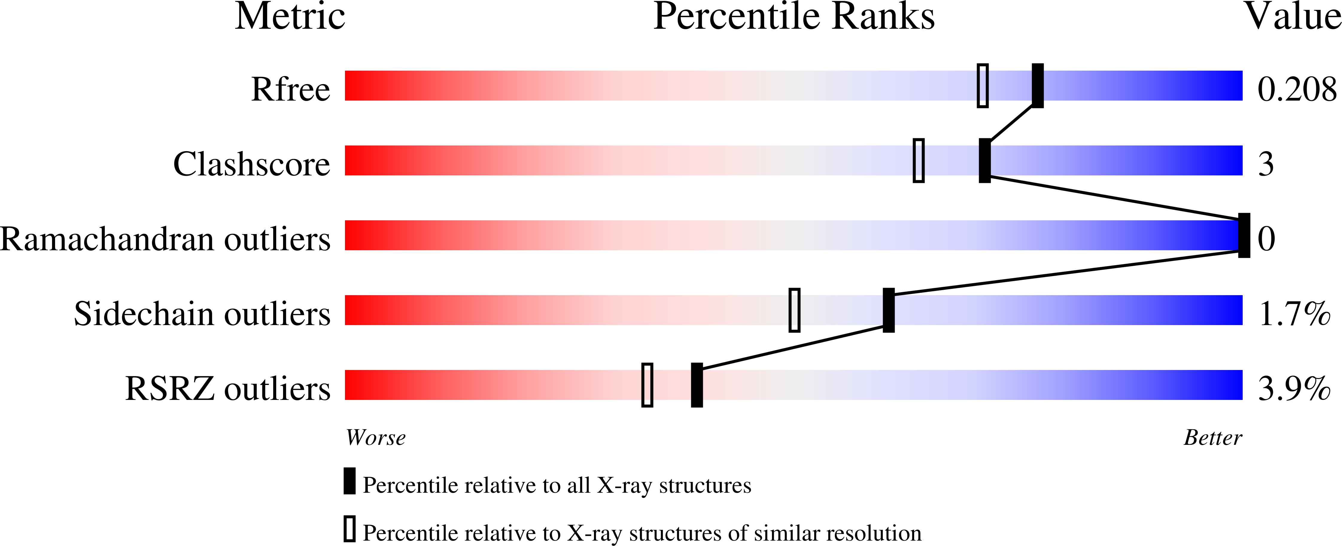
    6VYO, 6WKP, 7N3C, 7N3D, 7STR, 7STS, 7SUE

  • PubMed Abstract: 

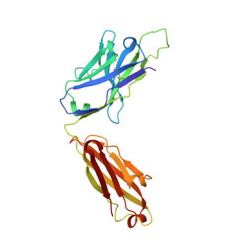
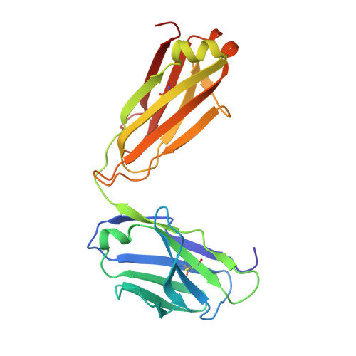
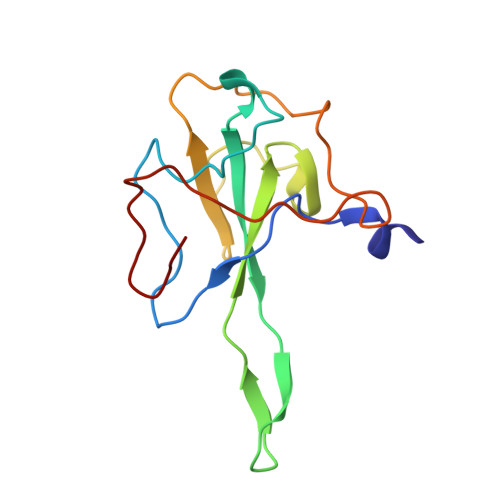
    Coronavirus nucleocapsid protein (NP) of SARS-CoV-2 plays a central role in many functions important for virus proliferation including packaging and protecting genomic RNA. The protein shares sequence, structure, and architecture with nucleocapsid proteins from betacoronaviruses. The N-terminal domain (NP RBD ) binds RNA and the C-terminal domain is responsible for dimerization. After infection, NP is highly expressed and triggers robust host immune response. The anti-NP antibodies are not protective and not neutralizing but can effectively detect viral proliferation soon after infection. Two structures of SARS-CoV-2 NP RBD were determined providing a continuous model from residue 48 to 173, including RNA binding region and key epitopes. Five structures of NP RBD complexes with human mAbs were isolated using an antigen-bait sorting. Complexes revealed a distinct complement-determining regions and unique sets of epitope recognition. This may assist in the early detection of pathogens and designing peptide-based vaccines. Mutations that significantly increase viral load were mapped on developed, full length NP model, likely impacting interactions with host proteins and viral RNA.


  • Organizational Affiliation

    Center for Structural Biology of Infectious Diseases, Consortium for Advanced Science and Engineering, University of Chicago, Chicago, IL 60367, USA.


Macromolecules
Find similar proteins by:  (by identity cutoff)  |  3D Structure
Entity ID: 1
MoleculeChains Sequence LengthOrganismDetailsImage
S24-202 Fab heavy chainA [auth H]227Homo sapiensMutation(s): 0 
Entity Groups  
Sequence Clusters30% Identity50% Identity70% Identity90% Identity95% Identity100% Identity
Sequence Annotations
Expand
  • Reference Sequence
Find similar proteins by:  (by identity cutoff)  |  3D Structure
Entity ID: 2
MoleculeChains Sequence LengthOrganismDetailsImage
S24-202 Fab light chainB [auth L]214Homo sapiensMutation(s): 0 
Entity Groups  
Sequence Clusters30% Identity50% Identity70% Identity90% Identity95% Identity100% Identity
Sequence Annotations
Expand
  • Reference Sequence
Find similar proteins by:  (by identity cutoff)  |  3D Structure
Entity ID: 3
MoleculeChains Sequence LengthOrganismDetailsImage
Nucleoprotein130Severe acute respiratory syndrome coronavirus 2Mutation(s): 0 
UniProt
Find proteins for P0DTC9 (Severe acute respiratory syndrome coronavirus 2)
Explore P0DTC9 
Go to UniProtKB:  P0DTC9
Entity Groups  
Sequence Clusters30% Identity50% Identity70% Identity90% Identity95% Identity100% Identity
UniProt GroupP0DTC9
Sequence Annotations
Expand
  • Reference Sequence
Small Molecules
Ligands 3 Unique
IDChains Name / Formula / InChI Key2D Diagram3D Interactions
IOD
Query on IOD

Download Ideal Coordinates CCD File 
I [auth H]
J [auth H]
K [auth H]
L [auth H]
O [auth L]
I [auth H],
J [auth H],
K [auth H],
L [auth H],
O [auth L],
P [auth L],
Q [auth L],
T [auth C],
U [auth C],
V [auth C],
W [auth C],
X [auth C]
IODIDE ION
I
XMBWDFGMSWQBCA-UHFFFAOYSA-M
PO4
Query on PO4

Download Ideal Coordinates CCD File 
Y [auth C]PHOSPHATE ION
O4 P
NBIIXXVUZAFLBC-UHFFFAOYSA-K
EDO
Query on EDO

Download Ideal Coordinates CCD File 
D [auth H]
E [auth H]
F [auth H]
G [auth H]
H
D [auth H],
E [auth H],
F [auth H],
G [auth H],
H,
M [auth L],
N [auth L],
R [auth C],
S [auth C]
1,2-ETHANEDIOL
C2 H6 O2
LYCAIKOWRPUZTN-UHFFFAOYSA-N
Experimental Data & Validation

Experimental Data

  • Method: X-RAY DIFFRACTION
  • Resolution: 1.82 Å
  • R-Value Free: 0.210 
  • R-Value Work: 0.179 
  • R-Value Observed: 0.181 
  • Space Group: P 1 21 1
Unit Cell:
Length ( Å )Angle ( ˚ )
a = 72.622α = 90
b = 64.02β = 91.091
c = 89.209γ = 90
Software Package:
Software NamePurpose
PHENIXrefinement
HKL-3000data reduction
HKL-3000data scaling
HKL-3000phasing
MOLREPphasing

Structure Validation

View Full Validation Report


Entry History & Funding Information

Deposition Data


Funding OrganizationLocationGrant Number
National Institutes of Health/National Institute Of Allergy and Infectious Diseases (NIH/NIAID)United States--

Revision History  (Full details and data files)

  • Version 1.0: 2021-07-07
    Type: Initial release
  • Version 1.1: 2021-07-28
    Changes: Database references, Structure summary
  • Version 1.2: 2023-10-18
    Changes: Data collection, Database references, Refinement description
  • Version 1.3: 2024-02-28
    Changes: Database references