5Z8X

Crystal structure of human LRRTM2


Experimental Data Snapshot

  • Method: X-RAY DIFFRACTION
  • Resolution: 3.15 Å
  • R-Value Free: 0.231 
  • R-Value Work: 0.203 
  • R-Value Observed: 0.204 

wwPDB Validation   3D Report Full Report


Ligand Structure Quality Assessment 


This is version 1.3 of the entry. See complete history


Literature

Structural insights into modulation and selectivity of transsynaptic neurexin-LRRTM interaction.

Yamagata, A.Goto-Ito, S.Sato, Y.Shiroshima, T.Maeda, A.Watanabe, M.Saitoh, T.Maenaka, K.Terada, T.Yoshida, T.Uemura, T.Fukai, S.

(2018) Nat Commun 9: 3964-3964

  • DOI: https://doi.org/10.1038/s41467-018-06333-8
  • Primary Citation of Related Structures:  
    5Z8X, 5Z8Y

  • PubMed Abstract: 

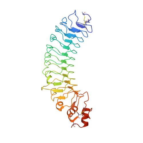
    Leucine-rich repeat transmembrane neuronal proteins (LRRTMs) function as postsynaptic organizers that induce excitatory synapses. Neurexins (Nrxns) and heparan sulfate proteoglycans have been identified as presynaptic ligands for LRRTMs. Specifically, LRRTM1 and LRRTM2 bind to the Nrxn splice variant lacking an insert at the splice site 4 (S4). Here, we report the crystal structure of the Nrxn1β-LRRTM2 complex at 3.4 Å resolution. The Nrxn1β-LRRTM2 interface involves Ca 2+ -mediated interactions and overlaps with the Nrxn-neuroligin interface. Together with structure-based mutational analyses at the molecular and cellular levels, the present structural analysis unveils the mechanism of selective binding between Nrxn and LRRTM1/2 and its modulation by the S4 insertion of Nrxn.


  • Organizational Affiliation

    Institute for Quantitative Biosciences, The University of Tokyo, Tokyo, 113-0032, Japan.


Macromolecules
Find similar proteins by:  (by identity cutoff)  |  3D Structure
Entity ID: 1
MoleculeChains Sequence LengthOrganismDetailsImage
Leucine-rich repeat transmembrane neuronal protein 2
A, B, C, D
346Homo sapiensMutation(s): 1 
Gene Names: LRRTM2KIAA0416LRRN2
UniProt & NIH Common Fund Data Resources
Find proteins for O43300 (Homo sapiens)
Explore O43300 
Go to UniProtKB:  O43300
PHAROS:  O43300
GTEx:  ENSG00000146006 
Entity Groups  
Sequence Clusters30% Identity50% Identity70% Identity90% Identity95% Identity100% Identity
UniProt GroupO43300
Sequence Annotations
Expand
  • Reference Sequence
Experimental Data & Validation

Experimental Data

  • Method: X-RAY DIFFRACTION
  • Resolution: 3.15 Å
  • R-Value Free: 0.231 
  • R-Value Work: 0.203 
  • R-Value Observed: 0.204 
  • Space Group: C 2 2 21
Unit Cell:
Length ( Å )Angle ( ˚ )
a = 90.691α = 90
b = 240.959β = 90
c = 259.434γ = 90
Software Package:
Software NamePurpose
PHENIXrefinement
HKL-2000data reduction
HKL-2000data scaling
MOLREPphasing

Structure Validation

View Full Validation Report


Ligand Structure Quality Assessment 


Entry History & Funding Information

Deposition Data


Funding OrganizationLocationGrant Number
JSPS/MEXTJapanJP16H04749
JSTJapanJPMJCR12M5

Revision History  (Full details and data files)

  • Version 1.0: 2018-10-10
    Type: Initial release
  • Version 1.1: 2018-10-31
    Changes: Data collection, Structure summary
  • Version 1.2: 2020-07-29
    Type: Remediation
    Reason: Carbohydrate remediation
    Changes: Data collection, Derived calculations, Structure summary
  • Version 1.3: 2023-11-22
    Changes: Data collection, Database references, Refinement description, Structure summary