4XBQ

Crystal Structure of Human Galectin-7 in Complex with Type 1 N-acetyllactosamine


Experimental Data Snapshot

  • Method: X-RAY DIFFRACTION
  • Resolution: 2.23 Å
  • R-Value Free: 0.225 
  • R-Value Work: 0.196 
  • R-Value Observed: 0.197 

wwPDB Validation   3D Report Full Report


This is version 2.1 of the entry. See complete history


Literature

Structural Basis Underlying the Binding Preference of Human Galectins-1, -3 and -7 for Gal beta 1-3/4GlcNAc.

Hsieh, T.J.Lin, H.Y.Tu, Z.Huang, B.S.Wu, S.C.Lin, C.H.

(2015) PLoS One 10: e0125946-e0125946

  • DOI: https://doi.org/10.1371/journal.pone.0125946
  • Primary Citation of Related Structures:  
    4XBL, 4XBN, 4XBQ

  • PubMed Abstract: 

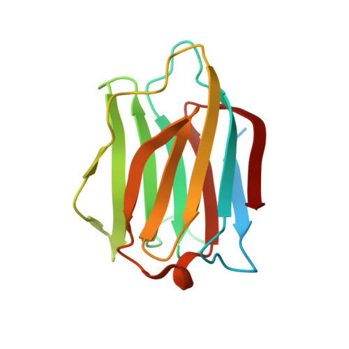
    Galectins represent β-galactoside-binding proteins and are known to bind Galβ1-3/4GlcNAc disaccharides (abbreviated as LN1 and LN2, respectively). Despite high sequence and structural homology shared by the carbohydrate recognition domain (CRD) of all galectin members, how each galectin displays different sugar-binding specificity still remains ambiguous. Herein we provided the first structural evidence of human galectins-1, 3-CRD and 7 in complex with LN1. Galectins-1 and 3 were shown to have higher affinity for LN2 than for LN1, while galectin-7 displayed the reversed specificity. In comparison with the previous LN2-complexed structures, the results indicated that the average glycosidic torsion angle of galectin-bound LN1 (ψ(LN1) ≈ 135°) was significantly differed from that of galectin-bound LN2 (ψ(LN2 )≈ -108°), i.e. the GlcNAc moiety adopted a different orientation to maintain essential interactions. Furthermore, we also identified an Arg-Asp/Glu-Glu-Arg salt-bridge network and the corresponding loop (to position the second Asp/Glu residue) critical for the LN1/2-binding preference.


  • Organizational Affiliation

    Institute of Biological Chemistry, Academia Sinica, Taipei, Taiwan.


Macromolecules
Find similar proteins by:  (by identity cutoff)  |  3D Structure
Entity ID: 1
MoleculeChains Sequence LengthOrganismDetailsImage
Galectin-7
A, B
155Homo sapiensMutation(s): 0 
Gene Names: LGALS7PIG1LGALS7B
UniProt & NIH Common Fund Data Resources
Find proteins for P47929 (Homo sapiens)
Explore P47929 
Go to UniProtKB:  P47929
PHAROS:  P47929
Entity Groups  
Sequence Clusters30% Identity50% Identity70% Identity90% Identity95% Identity100% Identity
UniProt GroupP47929
Sequence Annotations
Expand
  • Reference Sequence
Oligosaccharides

Help

Entity ID: 2
MoleculeChains Length2D Diagram Glycosylation3D Interactions
beta-D-galactopyranose-(1-3)-2-acetamido-2-deoxy-beta-D-glucopyranose
C, D
2N/A
Glycosylation Resources
GlyTouCan:  G00056MO
GlyCosmos:  G00056MO
GlyGen:  G00056MO
Experimental Data & Validation

Experimental Data

  • Method: X-RAY DIFFRACTION
  • Resolution: 2.23 Å
  • R-Value Free: 0.225 
  • R-Value Work: 0.196 
  • R-Value Observed: 0.197 
  • Space Group: P 21 21 21
Unit Cell:
Length ( Å )Angle ( ˚ )
a = 30.085α = 90
b = 55.411β = 90
c = 136.497γ = 90
Software Package:
Software NamePurpose
HKL-2000data collection
HKL-2000data processing
SCALEPACKdata scaling
PHASERphasing
PHENIXmodel building
PHENIXrefinement
PDB_EXTRACTdata extraction

Structure Validation

View Full Validation Report


Entry History & Funding Information

Deposition Data


Funding OrganizationLocationGrant Number
Academia SinicaTaiwanAS-022316
Ministry of Science and TechnologyTaiwan103-2113-M-001-023-MY3
Ministry of Science and TechnologyTaiwan102-2113-M-001-001-MY3
Ministry of Science and TechnologyTaiwan102-2923-M-001-001-MY3

Revision History  (Full details and data files)

  • Version 1.0: 2015-05-20
    Type: Initial release
  • Version 2.0: 2020-07-29
    Type: Remediation
    Reason: Carbohydrate remediation
    Changes: Atomic model, Data collection, Derived calculations, Source and taxonomy, Structure summary
  • Version 2.1: 2023-11-08
    Changes: Data collection, Database references, Derived calculations, Refinement description, Structure summary