3AIH

Human OS-9 MRH domain complexed with alpha3,alpha6-Man5


Experimental Data Snapshot

  • Method: X-RAY DIFFRACTION
  • Resolution: 2.10 Å
  • R-Value Free: 0.277 
  • R-Value Work: 0.228 
  • R-Value Observed: 0.230 

wwPDB Validation   3D Report Full Report


This is version 2.0 of the entry. See complete history


Literature

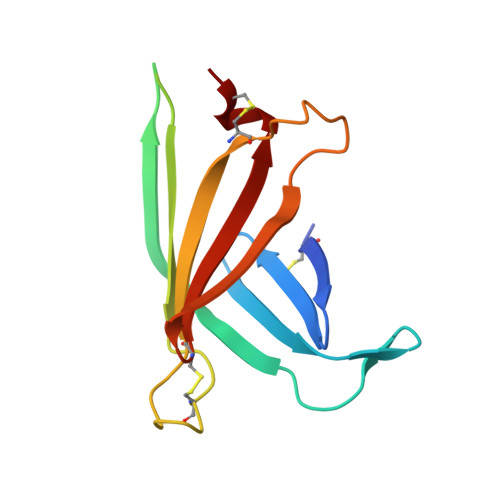
Structural Basis for Oligosaccharide Recognition of Misfolded Glycoproteins by OS-9 in ER-Associated Degradation

Satoh, T.Chen, Y.Hu, D.Hanashima, S.Yamamoto, K.Yamaguchi, Y.

(2010) Mol Cell 40: 905-916

  • DOI: https://doi.org/10.1016/j.molcel.2010.11.017
  • Primary Citation of Related Structures:  
    3AIH

  • PubMed Abstract: 

    Misfolded glycoproteins are translocated from endoplasmic reticulum (ER) into the cytosol for proteasome-mediated degradation. A mannose-6-phosphate receptor homology (MRH) domain is commonly identified in a variety of proteins and, in the case of OS-9 and XTP3-B, is involved in glycoprotein ER-associated degradation (ERAD). Trimming of outermost α1,2-linked mannose on C-arm of high-mannose-type glycan and binding of processed α1,6-linked mannosyl residues by the MRH domain are critical steps in guiding misfolded glycoproteins to enter ERAD. Here we report the crystal structure of a human OS-9 MRH domain (OS-9(MRH)) complexed with α3,α6-mannopentaose. The OS-9(MRH) has a flattened β-barrel structure with a characteristic P-type lectin fold and possesses distinctive double tryptophan residues in the oligosaccharide-binding site. Our crystallographic result in conjunction with nuclear magnetic resonance (NMR) spectroscopic and biochemical results provides structural insights into the mechanism whereby OS-9 specifically recognizes Manα1,6Manα1,6Man residues on the processed C-arm through the continuous double tryptophan (WW) motif.


  • Organizational Affiliation

    Structural Glycobiology Team, RIKEN Advanced Science Institute, Saitama 351-0198, Japan.


Macromolecules
Find similar proteins by:  (by identity cutoff)  |  3D Structure
Entity ID: 1
MoleculeChains Sequence LengthOrganismDetailsImage
Protein OS-9
A, B
124Homo sapiensMutation(s): 0 
Gene Names: OS9
UniProt & NIH Common Fund Data Resources
Find proteins for Q13438 (Homo sapiens)
Explore Q13438 
Go to UniProtKB:  Q13438
PHAROS:  Q13438
GTEx:  ENSG00000135506 
Entity Groups  
Sequence Clusters30% Identity50% Identity70% Identity90% Identity95% Identity100% Identity
UniProt GroupQ13438
Sequence Annotations
Expand
  • Reference Sequence
Oligosaccharides

Help

Entity ID: 2
MoleculeChains Length2D Diagram Glycosylation3D Interactions
alpha-D-mannopyranose-(1-6)-alpha-D-mannopyranose-(1-6)-beta-D-mannopyranose
C, D
3N/A
Glycosylation Resources
GlyTouCan:  G91725EU
GlyCosmos:  G91725EU
GlyGen:  G91725EU
Experimental Data & Validation

Experimental Data

  • Method: X-RAY DIFFRACTION
  • Resolution: 2.10 Å
  • R-Value Free: 0.277 
  • R-Value Work: 0.228 
  • R-Value Observed: 0.230 
  • Space Group: P 61 2 2
Unit Cell:
Length ( Å )Angle ( ˚ )
a = 73.1α = 90
b = 73.1β = 90
c = 180.3γ = 120
Software Package:
Software NamePurpose
HKL-2000data collection
SHELXSphasing
REFMACrefinement
DENZOdata reduction
SCALEPACKdata scaling

Structure Validation

View Full Validation Report


Entry History 

Deposition Data

Revision History  (Full details and data files)

  • Version 1.0: 2010-12-22
    Type: Initial release
  • Version 1.1: 2011-07-13
    Changes: Version format compliance
  • Version 1.2: 2014-12-17
    Changes: Atomic model, Derived calculations
  • Version 2.0: 2020-07-29
    Type: Remediation
    Reason: Carbohydrate remediation
    Changes: Advisory, Atomic model, Data collection, Database references, Derived calculations, Structure summary