6TSZ

The ULK4 Pseudokinase Domain Bound To ATPgammaS


Experimental Data Snapshot

  • Method: X-RAY DIFFRACTION
  • Resolution: 1.90 Å
  • R-Value Free: 0.253 
  • R-Value Work: 0.201 
  • R-Value Observed: 0.203 

wwPDB Validation   3D Report Full Report


Ligand Structure Quality Assessment 


This is version 1.3 of the entry. See complete history


Literature

Nucleotide Binding, Evolutionary Insights, and Interaction Partners of the Pseudokinase Unc-51-like Kinase 4.

Preuss, F.Chatterjee, D.Mathea, S.Shrestha, S.St-Germain, J.Saha, M.Kannan, N.Raught, B.Rottapel, R.Knapp, S.

(2020) Structure 28: 1184

  • DOI: https://doi.org/10.1016/j.str.2020.07.016
  • Primary Citation of Related Structures:  
    6TSZ

  • PubMed Abstract: 

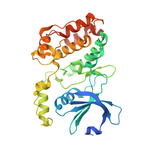
    Unc-51-like kinase 4 (ULK4) is a pseudokinase that has been linked to the development of several diseases. Even though sequence motifs required for ATP binding in kinases are lacking, ULK4 still tightly binds ATP and the presence of the co-factor is required for structural stability of ULK4. Here, we present a high-resolution structure of a ULK4-ATPγS complex revealing a highly unusual ATP binding mode in which the lack of the canonical VAIK motif lysine is compensated by K39, located N-terminal to αC. Evolutionary analysis suggests that degradation of active site motifs in metazoan ULK4 has co-occurred with an ULK4-specific activation loop, which stabilizes the C helix. In addition, cellular interaction studies using BioID and biochemical validation data revealed high confidence interactors of the pseudokinase and armadillo repeat domains. Many of the identified ULK4 interaction partners were centrosomal and tubulin-associated proteins and several active kinases suggesting interesting regulatory roles for ULK4.


  • Organizational Affiliation

    Institute for Pharmaceutical Chemistry, Johann Wolfgang Goethe-University, Max-von-Laue-Str. 9, 60438 Frankfurt am Main, Germany; Buchmann Institute for Molecular Life Sciences, Structural Genomics Consortium, Johann Wolfgang Goethe-University, Max-von-Laue-Str. 15, 60438 Frankfurt am Main, Germany.


Macromolecules
Find similar proteins by:  (by identity cutoff)  |  3D Structure
Entity ID: 1
MoleculeChains Sequence LengthOrganismDetailsImage
Serine/threonine-protein kinase ULK4A [auth U]305Homo sapiensMutation(s): 0 
Gene Names: ULK4
EC: 2.7.11.1
UniProt & NIH Common Fund Data Resources
Find proteins for Q96C45 (Homo sapiens)
Explore Q96C45 
Go to UniProtKB:  Q96C45
PHAROS:  Q96C45
GTEx:  ENSG00000168038 
Entity Groups  
Sequence Clusters30% Identity50% Identity70% Identity90% Identity95% Identity100% Identity
UniProt GroupQ96C45
Sequence Annotations
Expand
  • Reference Sequence
Small Molecules
Ligands 1 Unique
IDChains Name / Formula / InChI Key2D Diagram3D Interactions
AGS (Subject of Investigation/LOI)
Query on AGS

Download Ideal Coordinates CCD File 
B [auth U]PHOSPHOTHIOPHOSPHORIC ACID-ADENYLATE ESTER
C10 H16 N5 O12 P3 S
NLTUCYMLOPLUHL-KQYNXXCUSA-N
Experimental Data & Validation

Experimental Data

  • Method: X-RAY DIFFRACTION
  • Resolution: 1.90 Å
  • R-Value Free: 0.253 
  • R-Value Work: 0.201 
  • R-Value Observed: 0.203 
  • Space Group: P 1 21 1
Unit Cell:
Length ( Å )Angle ( ˚ )
a = 62.442α = 90
b = 41.468β = 113.063
c = 69.755γ = 90
Software Package:
Software NamePurpose
PHENIXrefinement
xia2data reduction
Aimlessdata scaling
PHASERphasing
Cootmodel building

Structure Validation

View Full Validation Report


Ligand Structure Quality Assessment 


Entry History 

Deposition Data

Revision History  (Full details and data files)

  • Version 1.0: 2020-01-01
    Type: Initial release
  • Version 1.1: 2020-09-02
    Changes: Database references, Source and taxonomy
  • Version 1.2: 2020-11-18
    Changes: Database references
  • Version 1.3: 2024-01-24
    Changes: Data collection, Database references, Refinement description