6NJO

Structure of the assembled ATPase EscN from the enteropathogenic E. coli (EPEC) type III secretion system


Experimental Data Snapshot

  • Method: ELECTRON MICROSCOPY
  • Resolution: 3.34 Å
  • Aggregation State: PARTICLE 
  • Reconstruction Method: SINGLE PARTICLE 

wwPDB Validation   3D Report Full Report


This is version 1.2 of the entry. See complete history


Literature

Cryo-EM structure of the homohexameric T3SS ATPase-central stalk complex reveals rotary ATPase-like asymmetry.

Majewski, D.D.Worrall, L.J.Hong, C.Atkinson, C.E.Vuckovic, M.Watanabe, N.Yu, Z.Strynadka, N.C.J.

(2019) Nat Commun 10: 626-626

  • DOI: https://doi.org/10.1038/s41467-019-08477-7
  • Primary Citation of Related Structures:  
    6NJO, 6NJP

  • PubMed Abstract: 

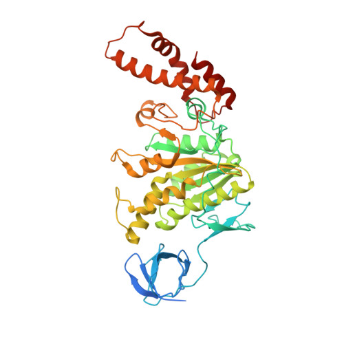
    Many Gram-negative bacteria, including causative agents of dysentery, plague, and typhoid fever, rely on a type III secretion system - a multi-membrane spanning syringe-like apparatus - for their pathogenicity. The cytosolic ATPase complex of this injectisome is proposed to play an important role in energizing secretion events and substrate recognition. We present the 3.3 Å resolution cryo-EM structure of the enteropathogenic Escherichia coli ATPase EscN in complex with its central stalk EscO. The structure shows an asymmetric pore with different functional states captured in its six catalytic sites, details directly supporting a rotary catalytic mechanism analogous to that of the heterohexameric F 1 /V 1 -ATPases despite its homohexameric nature. Situated at the C-terminal opening of the EscN pore is one molecule of EscO, with primary interaction mediated through an electrostatic interface. The EscN-EscO structure provides significant atomic insights into how the ATPase contributes to type III secretion, including torque generation and binding of chaperone/substrate complexes.


  • Organizational Affiliation

    Department of Biochemistry and Molecular Biology and the Center for Blood Research, University of British Columbia, Vancouver, BC, Canada.


Macromolecules
Find similar proteins by:  (by identity cutoff)  |  3D Structure
Entity ID: 1
MoleculeChains Sequence LengthOrganismDetailsImage
Translocator EscN
A, B, C, D, E
A, B, C, D, E, F
449Escherichia coli O127:H6 str. E2348/69Mutation(s): 0 
Gene Names: escNE2348C_3948
UniProt
Find proteins for B7UMA6 (Escherichia coli O127:H6 (strain E2348/69 / EPEC))
Explore B7UMA6 
Go to UniProtKB:  B7UMA6
Entity Groups  
Sequence Clusters30% Identity50% Identity70% Identity90% Identity95% Identity100% Identity
UniProt GroupB7UMA6
Sequence Annotations
Expand
  • Reference Sequence
Small Molecules
Ligands 3 Unique
IDChains Name / Formula / InChI Key2D Diagram3D Interactions
ADP
Query on ADP

Download Ideal Coordinates CCD File 
G [auth B],
J [auth C],
M [auth D],
P [auth E]
ADENOSINE-5'-DIPHOSPHATE
C10 H15 N5 O10 P2
XTWYTFMLZFPYCI-KQYNXXCUSA-N
AF3
Query on AF3

Download Ideal Coordinates CCD File 
I [auth B],
L [auth C],
O [auth D],
R [auth E]
ALUMINUM FLUORIDE
Al F3
KLZUFWVZNOTSEM-UHFFFAOYSA-K
MG
Query on MG

Download Ideal Coordinates CCD File 
H [auth B],
K [auth C],
N [auth D],
Q [auth E]
MAGNESIUM ION
Mg
JLVVSXFLKOJNIY-UHFFFAOYSA-N
Experimental Data & Validation

Experimental Data

  • Method: ELECTRON MICROSCOPY
  • Resolution: 3.34 Å
  • Aggregation State: PARTICLE 
  • Reconstruction Method: SINGLE PARTICLE 
EM Software:
TaskSoftware PackageVersion
RECONSTRUCTIONRELION3
MODEL REFINEMENTPHENIX

Structure Validation

View Full Validation Report


Entry History & Funding Information

Deposition Data


Funding OrganizationLocationGrant Number
Natural Sciences and Engineering Research Council (NSERC, Canada)Canada--
Canadian Institutes of Health Research (CIHR)Canada--
Howard Hughes Medical Institute (HHMI)United States--

Revision History  (Full details and data files)

  • Version 1.0: 2019-02-20
    Type: Initial release
  • Version 1.1: 2019-11-20
    Changes: Author supporting evidence
  • Version 1.2: 2024-03-20
    Changes: Data collection, Database references, Derived calculations, Refinement description