6FNZ

Crystal Structure of domain-swapped C-terminal domain of human doublecortin


Experimental Data Snapshot

  • Method: X-RAY DIFFRACTION
  • Resolution: 2.23 Å
  • R-Value Free: 0.231 
  • R-Value Work: 0.207 
  • R-Value Observed: 0.209 

wwPDB Validation   3D Report Full Report


This is version 1.3 of the entry. See complete history


Literature

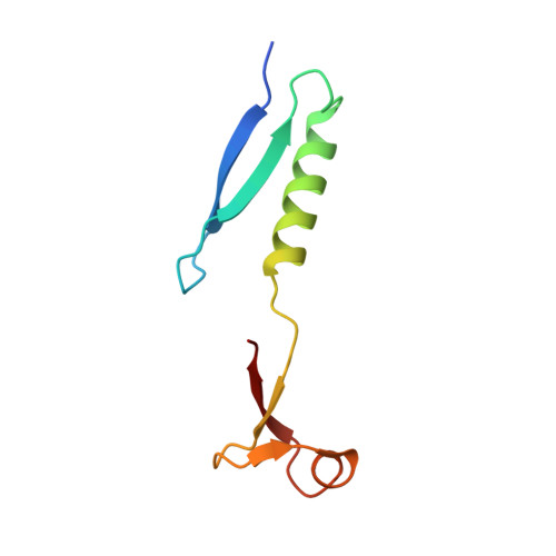
Domain swap in the C-terminal ubiquitin-like domain of human doublecortin.

Rufer, A.C.Kusznir, E.Burger, D.Stihle, M.Ruf, A.Rudolph, M.G.

(2018) Acta Crystallogr D Struct Biol 74: 450-462

  • DOI: https://doi.org/10.1107/S2059798318004813
  • Primary Citation of Related Structures:  
    6FNZ

  • PubMed Abstract: 

    Doublecortin, a microtubule-associated protein that is only produced during neurogenesis, cooperatively binds to microtubules and stimulates microtubule polymerization and cross-linking by unknown mechanisms. A domain swap is observed in the crystal structure of the C-terminal domain of doublecortin. As determined by analytical ultracentrifugation, an open conformation is also present in solution. At higher concentrations, higher-order oligomers of the domain are formed. The domain swap and additional interfaces observed in the crystal lattice can explain the formation of doublecortin tetramers or multimers, in line with the analytical ultracentrifugation data. Taken together, the domain swap offers a mechanism for the observed cooperative binding of doublecortin to microtubules. Doublecortin-induced cross-linking of microtubules can be explained by the same mechanism. The effect of several mutations leading to lissencephaly and double-cortex syndrome can be traced to the domain swap and the proposed self-association of doublecortin.


  • Organizational Affiliation

    pRED, Therapeutic Modalities, F. Hoffmann-La Roche, 4070 Basel, Switzerland.


Macromolecules
Find similar proteins by:  (by identity cutoff)  |  3D Structure
Entity ID: 1
MoleculeChains Sequence LengthOrganismDetailsImage
Neuronal migration protein doublecortin
A, B, C, D
81Homo sapiensMutation(s): 0 
Gene Names: DCXDBCNLISX
UniProt & NIH Common Fund Data Resources
Find proteins for O43602 (Homo sapiens)
Explore O43602 
Go to UniProtKB:  O43602
PHAROS:  O43602
GTEx:  ENSG00000077279 
Entity Groups  
Sequence Clusters30% Identity50% Identity70% Identity90% Identity95% Identity100% Identity
UniProt GroupO43602
Sequence Annotations
Expand
  • Reference Sequence

Find similar proteins by:  Sequence   |   3D Structure  

Entity ID: 2
MoleculeChains Sequence LengthOrganismDetailsImage
possible peptide
E, F, G
6unidentifiedMutation(s): 0 
Sequence Annotations
Expand
  • Reference Sequence
Experimental Data & Validation

Experimental Data

  • Method: X-RAY DIFFRACTION
  • Resolution: 2.23 Å
  • R-Value Free: 0.231 
  • R-Value Work: 0.207 
  • R-Value Observed: 0.209 
  • Space Group: P 43 21 2
Unit Cell:
Length ( Å )Angle ( ˚ )
a = 98.45α = 90
b = 98.45β = 90
c = 114.81γ = 90
Software Package:
Software NamePurpose
XSCALEdata scaling
PHENIXrefinement
PDB_EXTRACTdata extraction
XDSdata reduction
XSCALEdata scaling
PHASERphasing

Structure Validation

View Full Validation Report


Entry History 

Deposition Data

Revision History  (Full details and data files)

  • Version 1.0: 2018-04-04
    Type: Initial release
  • Version 1.1: 2018-05-09
    Changes: Data collection, Database references
  • Version 1.2: 2018-10-24
    Changes: Data collection, Derived calculations
  • Version 1.3: 2024-01-17
    Changes: Data collection, Database references, Refinement description