6VSJ

Cryo-electron microscopy structure of mouse coronavirus spike protein complexed with its murine receptor


Experimental Data Snapshot

  • Method: ELECTRON MICROSCOPY
  • Resolution: 3.94 Å
  • Aggregation State: PARTICLE 
  • Reconstruction Method: SINGLE PARTICLE 

wwPDB Validation   3D Report Full Report


This is version 1.3 of the entry. See complete history


Literature

Structure of mouse coronavirus spike protein complexed with receptor reveals mechanism for viral entry.

Shang, J.Wan, Y.Liu, C.Yount, B.Gully, K.Yang, Y.Auerbach, A.Peng, G.Baric, R.Li, F.

(2020) PLoS Pathog 16: e1008392-e1008392

  • DOI: https://doi.org/10.1371/journal.ppat.1008392
  • Primary Citation of Related Structures:  
    6VSJ

  • PubMed Abstract: 

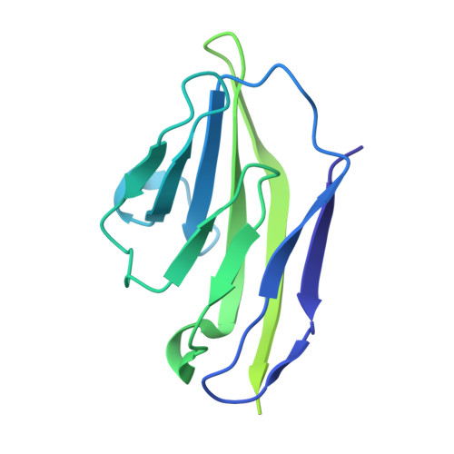
    Coronaviruses recognize a variety of receptors using different domains of their envelope-anchored spike protein. How these diverse receptor recognition patterns affect viral entry is unknown. Mouse hepatitis coronavirus (MHV) is the only known coronavirus that uses the N-terminal domain (NTD) of its spike to recognize a protein receptor, CEACAM1a. Here we determined the cryo-EM structure of MHV spike complexed with mouse CEACAM1a. The trimeric spike contains three receptor-binding S1 heads sitting on top of a trimeric membrane-fusion S2 stalk. Three receptor molecules bind to the sides of the spike trimer, where three NTDs are located. Receptor binding induces structural changes in the spike, weakening the interactions between S1 and S2. Using protease sensitivity and negative-stain EM analyses, we further showed that after protease treatment of the spike, receptor binding facilitated the dissociation of S1 from S2, allowing S2 to transition from pre-fusion to post-fusion conformation. Together these results reveal a new role of receptor binding in MHV entry: in addition to its well-characterized role in viral attachment to host cells, receptor binding also induces the conformational change of the spike and hence the fusion of viral and host membranes. Our study provides new mechanistic insight into coronavirus entry and highlights the diverse entry mechanisms used by different viruses.


  • Organizational Affiliation

    Department of Veterinary and Biomedical Sciences, University of Minnesota, Saint Paul, Minnesota, United States of America.


Macromolecules
Find similar proteins by:  (by identity cutoff)  |  3D Structure
Entity ID: 1
MoleculeChains Sequence LengthOrganismDetailsImage
Spike glycoproteinA,
C [auth B],
E [auth C]
1,275Murine hepatitis virus strain A59Mutation(s): 0 
Gene Names: S3
UniProt
Find proteins for P11224 (Murine coronavirus (strain A59))
Explore P11224 
Go to UniProtKB:  P11224
Entity Groups  
Sequence Clusters30% Identity50% Identity70% Identity90% Identity95% Identity100% Identity
UniProt GroupP11224
Sequence Annotations
Expand
  • Reference Sequence
Find similar proteins by:  (by identity cutoff)  |  3D Structure
Entity ID: 2
MoleculeChains Sequence LengthOrganismDetailsImage
Carcinoembryonic antigen-related cell adhesion molecule 1B [auth D],
D [auth E],
F
208Mus musculusMutation(s): 0 
Gene Names: Ceacam1BgpBgp1
UniProt
Find proteins for Q3LFS9 (Mus musculus)
Explore Q3LFS9 
Go to UniProtKB:  Q3LFS9
Entity Groups  
Sequence Clusters30% Identity50% Identity70% Identity90% Identity95% Identity100% Identity
UniProt GroupQ3LFS9
Sequence Annotations
Expand
  • Reference Sequence
Small Molecules
Ligands 1 Unique
IDChains Name / Formula / InChI Key2D Diagram3D Interactions
NAG
Query on NAG

Download Ideal Coordinates CCD File 
G [auth A]
H [auth A]
I [auth A]
J [auth D]
K [auth B]
G [auth A],
H [auth A],
I [auth A],
J [auth D],
K [auth B],
L [auth B],
M [auth B],
N [auth E],
O [auth C],
P [auth C],
Q [auth C],
R [auth F]
2-acetamido-2-deoxy-beta-D-glucopyranose
C8 H15 N O6
OVRNDRQMDRJTHS-FMDGEEDCSA-N
Experimental Data & Validation

Experimental Data

  • Method: ELECTRON MICROSCOPY
  • Resolution: 3.94 Å
  • Aggregation State: PARTICLE 
  • Reconstruction Method: SINGLE PARTICLE 

Structure Validation

View Full Validation Report


Entry History & Funding Information

Deposition Data


Funding OrganizationLocationGrant Number
National Institutes of Health/National Institute Of Allergy and Infectious Diseases (NIH/NIAID)United StatesR01AI089728

Revision History  (Full details and data files)

  • Version 1.0: 2020-03-04
    Type: Initial release
  • Version 1.1: 2020-03-18
    Changes: Structure summary
  • Version 1.2: 2020-03-25
    Changes: Database references
  • Version 1.3: 2020-07-29
    Type: Remediation
    Reason: Carbohydrate remediation
    Changes: Data collection, Derived calculations, Structure summary