6UET

SAM-bound SAM-IV riboswitch


Experimental Data Snapshot

  • Method: ELECTRON MICROSCOPY
  • Resolution: 4.10 Å
  • Aggregation State: PARTICLE 
  • Reconstruction Method: SINGLE PARTICLE 

wwPDB Validation   3D Report Full Report


This is version 1.1 of the entry. See complete history


Literature

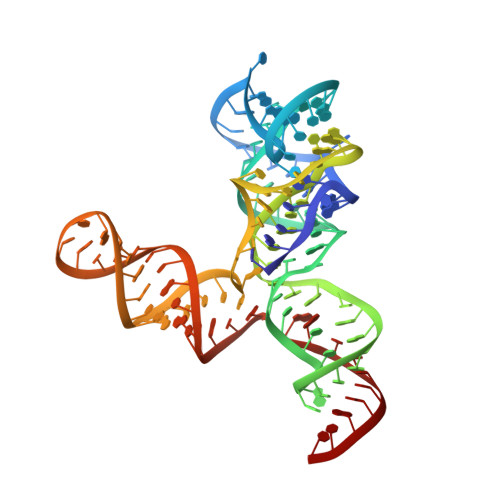
Cryo-EM structure of a 40 kDa SAM-IV riboswitch RNA at 3.7 angstrom resolution.

Zhang, K.Li, S.Kappel, K.Pintilie, G.Su, Z.Mou, T.C.Schmid, M.F.Das, R.Chiu, W.

(2019) Nat Commun 10: 5511-5511

  • DOI: https://doi.org/10.1038/s41467-019-13494-7
  • Primary Citation of Related Structures:  
    6UES, 6UET

  • PubMed Abstract: 

    Specimens below 50 kDa have generally been considered too small to be analyzed by single-particle cryo-electron microscopy (cryo-EM). The high flexibility of pure RNAs makes it difficult to obtain high-resolution structures by cryo-EM. In bacteria, riboswitches regulate sulfur metabolism through binding to the S-adenosylmethionine (SAM) ligand and offer compelling targets for new antibiotics. SAM-I, SAM-I/IV, and SAM-IV are the three most commonly found SAM riboswitches, but the structure of SAM-IV is still unknown. Here, we report the structures of apo and SAM-bound SAM-IV riboswitches (119-nt, ~40 kDa) to 3.7 Å and 4.1 Å resolution, respectively, using cryo-EM. The structures illustrate homologies in the ligand-binding core but distinct peripheral tertiary contacts in SAM-IV compared to SAM-I and SAM-I/IV. Our results demonstrate the feasibility of resolving small RNAs with enough detail to enable detection of their ligand-binding pockets and suggest that cryo-EM could play a role in structure-assisted drug design for RNA.


  • Organizational Affiliation

    Department of Bioengineering, and James H. Clark Center, Stanford University, Stanford, CA, 94305, USA.


Macromolecules
Find similar nucleic acids by:  (by identity cutoff)  |  3D Structure
Entity ID: 1
MoleculeChains LengthOrganismImage
RNA (119-MER)119Mycobacterium sp. MCS
Sequence Annotations
Expand
  • Reference Sequence
Small Molecules
Ligands 1 Unique
IDChains Name / Formula / InChI Key2D Diagram3D Interactions
SAM (Subject of Investigation/LOI)
Query on SAM

Download Ideal Coordinates CCD File 
B [auth A]S-ADENOSYLMETHIONINE
C15 H22 N6 O5 S
MEFKEPWMEQBLKI-FCKMPRQPSA-N
Experimental Data & Validation

Experimental Data

  • Method: ELECTRON MICROSCOPY
  • Resolution: 4.10 Å
  • Aggregation State: PARTICLE 
  • Reconstruction Method: SINGLE PARTICLE 
EM Software:
TaskSoftware PackageVersion
MODEL REFINEMENTPHENIX1.14
RECONSTRUCTIONcryoSPARC2

Structure Validation

View Full Validation Report


Entry History & Funding Information

Deposition Data


Funding OrganizationLocationGrant Number
National Institutes of Health/National Human Genome Research Institute (NIH/NHGRI)United StatesP41GM103832
National Institutes of Health/National Human Genome Research Institute (NIH/NHGRI)United StatesR01GM079429
National Institutes of Health/National Human Genome Research Institute (NIH/NHGRI)United StatesS10OD021600

Revision History  (Full details and data files)

  • Version 1.0: 2019-12-18
    Type: Initial release
  • Version 1.1: 2024-03-20
    Changes: Data collection, Database references