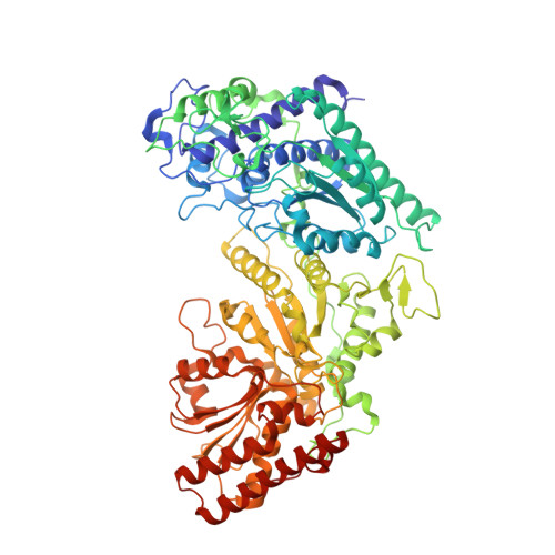
6GUA

Xylulose 5-phosphate phosphoketolase from Lactococcus lactis


Experimental Data Snapshot

  • Method: X-RAY DIFFRACTION
  • Resolution: 1.95 Å
  • R-Value Free: 0.261 
  • R-Value Work: 0.212 

  • Method: SOLUTION SCATTERING

wwPDB Validation   3D Report Full Report


Ligand Structure Quality Assessment 


This is version 1.2 of the entry. See complete history


Literature

Crystal structure of a xylulose 5-phosphate phosphoketolase. Insights into the substrate specificity for xylulose 5-phosphate.

Scheidig, A.J.Horvath, D.Szedlacsek, S.E.

(2019) J Struct Biol 207: 85-102

  • DOI: https://doi.org/10.1016/j.jsb.2019.04.017
  • Primary Citation of Related Structures:  
    6GUA

  • PubMed Abstract: 

    Phosphoketolases (PK) are TPP-dependent enzymes which play essential roles in carbohydrate metabolism of numerous bacteria. Depending on the substrate specificity PKs can be subdivided into xylulose 5-phosphate (X5P) specific PKs (XPKs) and PKs which accept both X5P and fructose 6-phosphate (F6P) (XFPKs). Despite their key metabolic importance, so far only the crystal structures of two XFPKs have been reported. There are no reported structures for any XPKs and for any complexes between PK and substrate. One of the major unknowns concerning PKs mechanism of action is related to the structural determinants of PKs substrate specificity for X5P or F6P. We report here the crystal structure of XPK from Lactococcus lactis (XPK-Ll) at 2.1 Å resolution. Using small angle X-ray scattering (SAXS) we proved that XPK-Ll is a dimer in solution. Towards better understanding of PKs substrate specificity, we performed flexible docking of TPP-X5P and TPP-F6P on crystal structures of XPK-Ll, two XFPKs and transketolase (TK). Calculated structure-based binding energies consistently support XPK-Ll preference for X5P. Analysis of structural models thus obtained show that substrates adopt moderately different conformation in PKs active sites following distinct networks of polar interactions. Based on the here reported structure of XPK-Ll we propose the most probable amino acid residues involved in the catalytic steps of reaction mechanism. Altogether our results suggest that PKs substrate preference for X5P or F6P is the outcome of a fine balance between specific binding network and dissimilar catalytic residues depending on the enzyme (XPK or XFPK) - substrate (X5P or F6P) couples.


  • Organizational Affiliation

    Structural Biology, Zoological Institute, Kiel University, Am Botanischen Garten 1-9, 24118 Kiel, Germany. Electronic address: axel.scheidig@strubio.uni-kiel.de.


Macromolecules
Find similar proteins by:  (by identity cutoff)  |  3D Structure
Entity ID: 1
MoleculeChains Sequence LengthOrganismDetailsImage
Probable phosphoketolase
A, B, C, D, E
A, B, C, D, E, F, G, H
822Lactococcus lactis subsp. lactis Il1403Mutation(s): 0 
Gene Names: LL1502L138230
EC: 4.1.2
UniProt
Find proteins for Q9CFH4 (Lactococcus lactis subsp. lactis (strain IL1403))
Explore Q9CFH4 
Go to UniProtKB:  Q9CFH4
Entity Groups  
Sequence Clusters30% Identity50% Identity70% Identity90% Identity95% Identity100% Identity
UniProt GroupQ9CFH4
Sequence Annotations
Expand
  • Reference Sequence
Small Molecules
Ligands 3 Unique
IDChains Name / Formula / InChI Key2D Diagram3D Interactions
TPP
Query on TPP

Download Ideal Coordinates CCD File 
AA [auth G]
DA [auth H]
I [auth A]
M [auth B]
O [auth C]
AA [auth G],
DA [auth H],
I [auth A],
M [auth B],
O [auth C],
S [auth D],
U [auth E],
Y [auth F]
THIAMINE DIPHOSPHATE
C12 H19 N4 O7 P2 S
AYEKOFBPNLCAJY-UHFFFAOYSA-O
PO4
Query on PO4

Download Ideal Coordinates CCD File 
CA [auth G]
FA [auth H]
K [auth A]
L [auth B]
Q [auth C]
CA [auth G],
FA [auth H],
K [auth A],
L [auth B],
Q [auth C],
R [auth C],
W [auth E],
X [auth E]
PHOSPHATE ION
O4 P
NBIIXXVUZAFLBC-UHFFFAOYSA-K
MG
Query on MG

Download Ideal Coordinates CCD File 
BA [auth G]
EA [auth H]
J [auth A]
N [auth B]
P [auth C]
BA [auth G],
EA [auth H],
J [auth A],
N [auth B],
P [auth C],
T [auth D],
V [auth E],
Z [auth F]
MAGNESIUM ION
Mg
JLVVSXFLKOJNIY-UHFFFAOYSA-N
Experimental Data & Validation

Experimental Data

  • Method: X-RAY DIFFRACTION
  • Resolution: 1.95 Å
  • R-Value Free: 0.261 
  • R-Value Work: 0.212 
  • Space Group: P 1 21 1
Unit Cell:
Length ( Å )Angle ( ˚ )
a = 158.1α = 90
b = 141.5β = 90.29
c = 161.15γ = 90
Software Package:
Software NamePurpose
PHENIXrefinement
XDSdata reduction
XSCALEdata scaling
MOLREPphasing

Structure Validation

View Full Validation Report


Ligand Structure Quality Assessment 


Entry History 

Deposition Data

Revision History  (Full details and data files)

  • Version 1.0: 2019-05-22
    Type: Initial release
  • Version 1.1: 2019-06-19
    Changes: Data collection, Database references
  • Version 1.2: 2024-01-17
    Changes: Data collection, Database references, Derived calculations, Refinement description