6CE9

Insulin Receptor ectodomain in complex with two insulin molecules


Experimental Data Snapshot

  • Method: ELECTRON MICROSCOPY
  • Resolution: 4.30 Å
  • Aggregation State: PARTICLE 
  • Reconstruction Method: SINGLE PARTICLE 

wwPDB Validation   3D Report Full Report


This is version 2.0 of the entry. See complete history


Literature

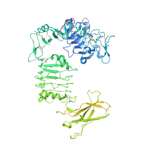
Structure of the insulin receptor-insulin complex by single-particle cryo-EM analysis.

Scapin, G.Dandey, V.P.Zhang, Z.Prosise, W.Hruza, A.Kelly, T.Mayhood, T.Strickland, C.Potter, C.S.Carragher, B.

(2018) Nature 556: 122-125

  • DOI: https://doi.org/10.1038/nature26153
  • Primary Citation of Related Structures:  
    6CE7, 6CE9, 6CEB

  • PubMed Abstract: 

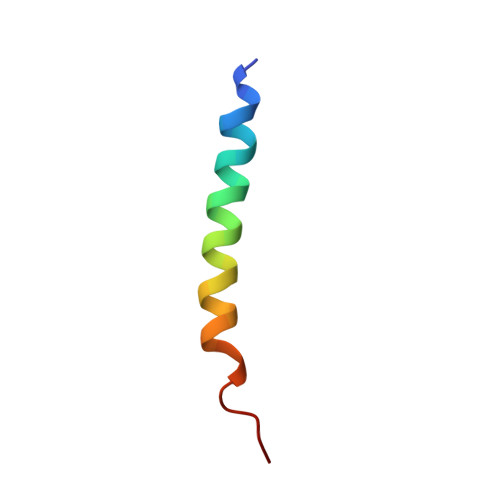
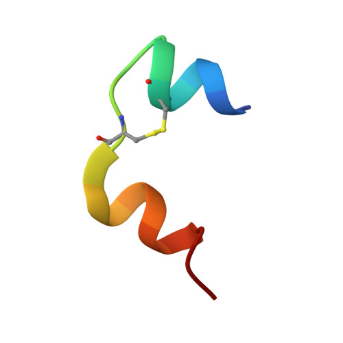
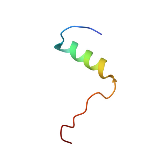
    The insulin receptor is a dimeric protein that has a crucial role in controlling glucose homeostasis, regulating lipid, protein and carbohydrate metabolism, and modulating brain neurotransmitter levels. Insulin receptor dysfunction has been associated with many diseases, including diabetes, cancer and Alzheimer's disease. The primary sequence of the receptor has been known since the 1980s, and is composed of an extracellular portion (the ectodomain, ECD), a single transmembrane helix and an intracellular tyrosine kinase domain. Binding of insulin to the dimeric ECD triggers auto-phosphorylation of the tyrosine kinase domain and subsequent activation of downstream signalling molecules. Biochemical and mutagenesis data have identified two putative insulin-binding sites, S1 and S2. The structures of insulin bound to an ECD fragment containing S1 and of the apo ectodomain have previously been reported, but details of insulin binding to the full receptor and the signal propagation mechanism are still not understood. Here we report single-particle cryo-electron microscopy reconstructions of the 1:2 (4.3 Å) and 1:1 (7.4 Å) complexes of the insulin receptor ECD dimer with insulin. The symmetrical 4.3 Å structure shows two insulin molecules per dimer, each bound between the leucine-rich subdomain L1 of one monomer and the first fibronectin-like domain (FnIII-1) of the other monomer, and making extensive interactions with the α-subunit C-terminal helix (α-CT helix). The 7.4 Å structure has only one similarly bound insulin per receptor dimer. The structures confirm the binding interactions at S1 and define the full S2 binding site. These insulin receptor states suggest that recruitment of the α-CT helix upon binding of the first insulin changes the relative subdomain orientations and triggers downstream signal propagation.


  • Organizational Affiliation

    Merck & Co., Department of Biochemical Engineering & Structure, 2000 Galloping Hill Road, Kenilworth, New Jersey 07033, USA.


Macromolecules
Find similar proteins by:  (by identity cutoff)  |  3D Structure
Entity ID: 1
MoleculeChains Sequence LengthOrganismDetailsImage
Insulin receptor
A, B
917Homo sapiensMutation(s): 0 
Gene Names: INSR
EC: 2.7.10.1
UniProt & NIH Common Fund Data Resources
Find proteins for P06213 (Homo sapiens)
Explore P06213 
Go to UniProtKB:  P06213
PHAROS:  P06213
GTEx:  ENSG00000171105 
Entity Groups  
Sequence Clusters30% Identity50% Identity70% Identity90% Identity95% Identity100% Identity
UniProt GroupP06213
Sequence Annotations
Expand
  • Reference Sequence
Find similar proteins by:  (by identity cutoff)  |  3D Structure
Entity ID: 2
MoleculeChains Sequence LengthOrganismDetailsImage
Insulin receptorC [auth M],
H [auth P]
30Homo sapiensMutation(s): 0 
Gene Names: INSR
EC: 2.7.10.1
UniProt & NIH Common Fund Data Resources
Find proteins for P06213 (Homo sapiens)
Explore P06213 
Go to UniProtKB:  P06213
PHAROS:  P06213
GTEx:  ENSG00000171105 
Entity Groups  
Sequence Clusters30% Identity50% Identity70% Identity90% Identity95% Identity100% Identity
UniProt GroupP06213
Sequence Annotations
Expand
  • Reference Sequence

Find similar proteins by:  Sequence   |   3D Structure  

Entity ID: 3
MoleculeChains Sequence LengthOrganismDetailsImage
Insulin A chainD [auth K],
F [auth N]
21Homo sapiensMutation(s): 0 
UniProt & NIH Common Fund Data Resources
Find proteins for P01308 (Homo sapiens)
Explore P01308 
Go to UniProtKB:  P01308
PHAROS:  P01308
GTEx:  ENSG00000254647 
Entity Groups  
Sequence Clusters30% Identity50% Identity70% Identity90% Identity95% Identity100% Identity
UniProt GroupP01308
Sequence Annotations
Expand
  • Reference Sequence
Find similar proteins by:  (by identity cutoff)  |  3D Structure
Entity ID: 4
MoleculeChains Sequence LengthOrganismDetailsImage
Insulin B chainE [auth L],
G [auth O]
30Ovis ariesMutation(s): 0 
UniProt
Find proteins for P01318 (Ovis aries)
Explore P01318 
Go to UniProtKB:  P01318
Entity Groups  
Sequence Clusters30% Identity50% Identity70% Identity90% Identity95% Identity100% Identity
UniProt GroupP01318
Sequence Annotations
Expand
  • Reference Sequence
Oligosaccharides

Help

Entity ID: 5
MoleculeChains Length2D Diagram Glycosylation3D Interactions
2-acetamido-2-deoxy-beta-D-glucopyranose-(1-4)-[alpha-L-fucopyranose-(1-6)]2-acetamido-2-deoxy-beta-D-glucopyranoseI [auth C],
K [auth E],
N [auth H],
P [auth J]
3N-Glycosylation
Glycosylation Resources
GlyTouCan:  G21290RB
GlyCosmos:  G21290RB
GlyGen:  G21290RB
Entity ID: 6
MoleculeChains Length2D Diagram Glycosylation3D Interactions
beta-D-mannopyranose-(1-4)-2-acetamido-2-deoxy-beta-D-glucopyranose-(1-4)-2-acetamido-2-deoxy-beta-D-glucopyranoseJ [auth D],
O [auth I]
3N-Glycosylation
Glycosylation Resources
GlyTouCan:  G15407YE
GlyCosmos:  G15407YE
GlyGen:  G15407YE
Entity ID: 7
MoleculeChains Length2D Diagram Glycosylation3D Interactions
2-acetamido-2-deoxy-beta-D-glucopyranose-(1-4)-2-acetamido-2-deoxy-beta-D-glucopyranoseL [auth F],
Q
2N-Glycosylation
Glycosylation Resources
GlyTouCan:  G42666HT
GlyCosmos:  G42666HT
GlyGen:  G42666HT
Entity ID: 8
MoleculeChains Length2D Diagram Glycosylation3D Interactions
alpha-L-fucopyranose-(1-6)-2-acetamido-2-deoxy-beta-D-glucopyranoseM [auth G],
R
2N-Glycosylation
Glycosylation Resources
GlyTouCan:  G86851RC
GlyCosmos:  G86851RC
GlyGen:  G86851RC
Experimental Data & Validation

Experimental Data

  • Method: ELECTRON MICROSCOPY
  • Resolution: 4.30 Å
  • Aggregation State: PARTICLE 
  • Reconstruction Method: SINGLE PARTICLE 
EM Software:
TaskSoftware PackageVersion
MODEL REFINEMENTPHENIX
RECONSTRUCTIONcryoSPARC0.6.5

Structure Validation

View Full Validation Report


Entry History & Funding Information

Deposition Data


Funding OrganizationLocationGrant Number
National Institutes of Health/National Institute of General Medical Sciences (NIH/NIGMS)United StatesGM103310
National Institutes of Health/National Institute on Deafness and Other Communication Disorders (NIH/NIDCD)United StatesOD019994
Simons FoundationUnited States349247

Revision History  (Full details and data files)

  • Version 1.0: 2018-03-14
    Type: Initial release
  • Version 1.1: 2018-03-21
    Changes: Database references
  • Version 1.2: 2018-04-18
    Changes: Data collection, Database references
  • Version 1.3: 2019-12-18
    Changes: Author supporting evidence, Data collection, Other
  • Version 2.0: 2020-07-29
    Type: Remediation
    Reason: Carbohydrate remediation
    Changes: Advisory, Atomic model, Data collection, Derived calculations, Structure summary