Electrostatic Interactions between Hendra Virus Matrix Proteins Are Required for Efficient Virus-Like-Particle Assembly.
Liu, Y.C., Grusovin, J., Adams, T.E.(2018) J Virol 92
- PubMed: 29695428
- DOI: https://doi.org/10.1128/JVI.00143-18
- Primary Citation of Related Structures:
6BK6 - PubMed Abstract:
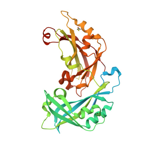
Hendra virus (HeV) is a zoonotic paramyxovirus belonging to the genus Henipavirus HeV is highly pathogenic, and it can cause severe neurological and respiratory illnesses in both humans and animals, with an extremely high mortality rate of up to 70%. Among the genes that HeV encodes, the matrix (M) protein forms an integral part of the virion structure and plays critical roles in coordinating viral assembly and budding. Nevertheless, the molecular mechanism of this process is not fully elucidated. Here, we determined the crystal structure of HeV M to 2.5-Å resolution. The dimeric structural configuration of HeV M is similar to that of Newcastle disease virus (NDV) M and is fundamental to protein stability and effective virus-like-particle (VLP) formation. Analysis of the crystal packing revealed a notable interface between the α1 and α2 helices of neighboring HeV M dimers, with key residues sharing degrees of sequence conservation among henipavirus M proteins. Structurally, a network of electrostatic interactions dominates the α1-α2 interactions, involving residues Arg57 from the α1 helix and Asp105 and Glu108 from the α2 helix. The disruption of the α1-α2 interactions using engineered charge reversal substitutions (R57E, R57D, and E108R) resulted in significant reduction or abrogation of VLP production. This phenotype was reversible with an R57E E108R mutant that was designed to partly restore salt bridge contacts. Collectively, our results define and validate previously underappreciated regions of henipavirus M proteins that are crucial for productive VLP assembly. IMPORTANCE Hendra virus is a henipavirus associated with lethal infections in humans. It is classified as a biosafety level 4 (BSL4) agent, and there are currently no preventive or therapeutic treatments available against HeV. Vital to henipavirus pathogenesis, the structural protein M has been implicated in viral assembly and budding, as well as host-virus interactions. However, there is no structural information available for henipavirus M, and the basis of M-driven viral assembly is not fully elucidated. We demonstrate the first three-dimensional structure of a henipavirus M protein. We show the dimeric organization of HeV M as a basic unit for higher-order oligomerization. Additionally, we define key regions/residues of HeV M that are required for productive virus-like-particle formation. These findings provide the first insight into the mechanism of M-driven assembly in henipavirus.
Organizational Affiliation:
CSIRO Manufacturing, Parkville, Victoria, Australia john.liu@csiro.au.