6B3R

Structure of the mechanosensitive channel Piezo1


Experimental Data Snapshot

  • Method: ELECTRON MICROSCOPY
  • Resolution: 3.80 Å
  • Aggregation State: PARTICLE 
  • Reconstruction Method: SINGLE PARTICLE 

wwPDB Validation   3D Report Full Report


This is version 1.1 of the entry. See complete history


Literature

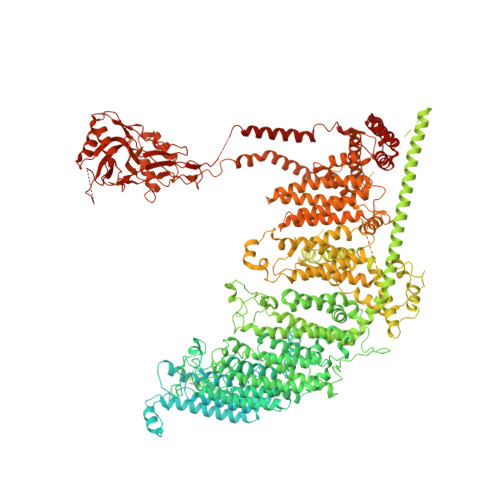
Structure-based membrane dome mechanism for Piezo mechanosensitivity.

Guo, Y.R.MacKinnon, R.

(2017) Elife 6

  • DOI: https://doi.org/10.7554/eLife.33660
  • Primary Citation of Related Structures:  
    6B3R

  • PubMed Abstract: 

    Mechanosensitive ion channels convert external mechanical stimuli into electrochemical signals for critical processes including touch sensation, balance, and cardiovascular regulation. The best understood mechanosensitive channel, MscL, opens a wide pore, which accounts for mechanosensitive gating due to in-plane area expansion. Eukaryotic Piezo channels have a narrow pore and therefore must capture mechanical forces to control gating in another way. We present a cryo-EM structure of mouse Piezo1 in a closed conformation at 3.7Å-resolution. The channel is a triskelion with arms consisting of repeated arrays of 4-TM structural units surrounding a pore. Its shape deforms the membrane locally into a dome. We present a hypothesis in which the membrane deformation changes upon channel opening. Quantitatively, membrane tension will alter gating energetics in proportion to the change in projected area under the dome. This mechanism can account for highly sensitive mechanical gating in the setting of a narrow, cation-selective pore.


  • Organizational Affiliation

    Laboratory of Molecular Neurobiology and Biophysics, Howard Hughes Medical Institute, The Rockefeller University, New York, United States.


Macromolecules
Find similar proteins by:  (by identity cutoff)  |  3D Structure
Entity ID: 1
MoleculeChains Sequence LengthOrganismDetailsImage
Piezo-type mechanosensitive ion channel component 1A,
C,
D [auth E]
2,547Mus musculusMutation(s): 0 
Gene Names: Piezo1Fam38a
Membrane Entity: Yes 
UniProt & NIH Common Fund Data Resources
Find proteins for E2JF22 (Mus musculus)
Explore E2JF22 
Go to UniProtKB:  E2JF22
IMPC:  MGI:3603204
Entity Groups  
Sequence Clusters30% Identity50% Identity70% Identity90% Identity95% Identity100% Identity
UniProt GroupE2JF22
Sequence Annotations
Expand
  • Reference Sequence

Find similar proteins by:  Sequence   |   3D Structure  

Entity ID: 2
MoleculeChains Sequence LengthOrganismDetailsImage
Piezo-type mechanosensitive ion channel component 1, unknown fragmentB,
E [auth D],
F
16Mus musculusMutation(s): 0 
Gene Names: Piezo1
Sequence Annotations
Expand
  • Reference Sequence
Experimental Data & Validation

Experimental Data

  • Method: ELECTRON MICROSCOPY
  • Resolution: 3.80 Å
  • Aggregation State: PARTICLE 
  • Reconstruction Method: SINGLE PARTICLE 
EM Software:
TaskSoftware PackageVersion
RECONSTRUCTIONRELION2
MODEL REFINEMENTPHENIX1.12-2829

Structure Validation

View Full Validation Report


Entry History 

Deposition Data

Revision History  (Full details and data files)

  • Version 1.0: 2017-12-20
    Type: Initial release
  • Version 1.1: 2018-05-16
    Changes: Data collection, Database references