5W0P

Crystal structure of rhodopsin bound to visual arrestin determined by X-ray free electron laser


Experimental Data Snapshot

  • Method: X-RAY DIFFRACTION
  • Resolution: 3.01 Å
  • R-Value Free: 0.272 
  • R-Value Work: 0.234 
  • R-Value Observed: 0.237 

wwPDB Validation   3D Report Full Report


This is version 2.1 of the entry. See complete history


Literature

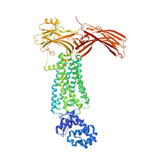
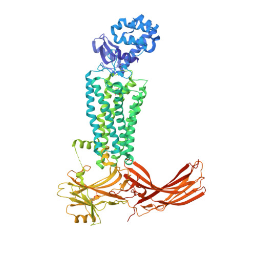
Identification of Phosphorylation Codes for Arrestin Recruitment by G Protein-Coupled Receptors.

Zhou, X.E.He, Y.de Waal, P.W.Gao, X.Kang, Y.Van Eps, N.Yin, Y.Pal, K.Goswami, D.White, T.A.Barty, A.Latorraca, N.R.Chapman, H.N.Hubbell, W.L.Dror, R.O.Stevens, R.C.Cherezov, V.Gurevich, V.V.Griffin, P.R.Ernst, O.P.Melcher, K.Xu, H.E.

(2017) Cell 170: 457-469.e13

  • DOI: https://doi.org/10.1016/j.cell.2017.07.002
  • Primary Citation of Related Structures:  
    5W0P

  • PubMed Abstract: 

    G protein-coupled receptors (GPCRs) mediate diverse signaling in part through interaction with arrestins, whose binding promotes receptor internalization and signaling through G protein-independent pathways. High-affinity arrestin binding requires receptor phosphorylation, often at the receptor's C-terminal tail. Here, we report an X-ray free electron laser (XFEL) crystal structure of the rhodopsin-arrestin complex, in which the phosphorylated C terminus of rhodopsin forms an extended intermolecular β sheet with the N-terminal β strands of arrestin. Phosphorylation was detected at rhodopsin C-terminal tail residues T336 and S338. These two phospho-residues, together with E341, form an extensive network of electrostatic interactions with three positively charged pockets in arrestin in a mode that resembles binding of the phosphorylated vasopressin-2 receptor tail to β-arrestin-1. Based on these observations, we derived and validated a set of phosphorylation codes that serve as a common mechanism for phosphorylation-dependent recruitment of arrestins by GPCRs.


  • Organizational Affiliation

    VARI-SIMM Center, Center for Structure and Function of Drug Targets, CAS-Key Laboratory of Receptor Research, Shanghai Institute of Materia Medica, Chinese Academy of Sciences, Shanghai 201203, China; Laboratory of Structural Sciences, Center for Structural Biology and Drug Discovery, Van Andel Research Institute, Grand Rapids, MI 49503, USA.


Macromolecules
Find similar proteins by:  (by identity cutoff)  |  3D Structure
Entity ID: 1
MoleculeChains Sequence LengthOrganismDetailsImage
Endolysin,Rhodopsin,S-arrestin
A, B
906Enterobacteria phage RB55Homo sapiensMus musculus
This entity is chimeric
Mutation(s): 9 
Gene Names: eRB55_p125RHOOPN2Sag
EC: 3.2.1.17
Membrane Entity: Yes 
UniProt & NIH Common Fund Data Resources
Find proteins for P00720 (Enterobacteria phage T4)
Explore P00720 
Go to UniProtKB:  P00720
Find proteins for P08100 (Homo sapiens)
Explore P08100 
Go to UniProtKB:  P08100
PHAROS:  P08100
GTEx:  ENSG00000163914 
Find proteins for P20443 (Mus musculus)
Explore P20443 
Go to UniProtKB:  P20443
IMPC:  MGI:98227
Entity Groups  
Sequence Clusters30% Identity50% Identity70% Identity90% Identity95% Identity100% Identity
UniProt GroupsP00720P08100P20443
Sequence Annotations
Expand
  • Reference Sequence
Find similar proteins by:  (by identity cutoff)  |  3D Structure
Entity ID: 2
MoleculeChains Sequence LengthOrganismDetailsImage
Endolysin,Rhodopsin,S-arrestin
C, D
906Enterobacteria phage RB55Homo sapiensMus musculus
This entity is chimeric
Mutation(s): 9 
Gene Names: eRB55_p125RHOOPN2Sag
EC: 3.2.1.17
Membrane Entity: Yes 
UniProt & NIH Common Fund Data Resources
Find proteins for P00720 (Enterobacteria phage T4)
Explore P00720 
Go to UniProtKB:  P00720
Find proteins for P08100 (Homo sapiens)
Explore P08100 
Go to UniProtKB:  P08100
PHAROS:  P08100
GTEx:  ENSG00000163914 
Find proteins for P20443 (Mus musculus)
Explore P20443 
Go to UniProtKB:  P20443
IMPC:  MGI:98227
Entity Groups  
Sequence Clusters30% Identity50% Identity70% Identity90% Identity95% Identity100% Identity
UniProt GroupsP00720P08100P20443
Sequence Annotations
Expand
  • Reference Sequence
Oligosaccharides

Help

Entity ID: 3
MoleculeChains Length2D Diagram Glycosylation3D Interactions
2-acetamido-2-deoxy-beta-D-glucopyranose-(1-4)-2-acetamido-2-deoxy-beta-D-glucopyranose
E, F, G, H
2N-Glycosylation
Glycosylation Resources
GlyTouCan:  G42666HT
GlyCosmos:  G42666HT
GlyGen:  G42666HT
Small Molecules
Modified Residues  2 Unique
IDChains TypeFormula2D DiagramParent
SEP
Query on SEP
A, B
L-PEPTIDE LINKINGC3 H8 N O6 PSER
TPO
Query on TPO
A, B
L-PEPTIDE LINKINGC4 H10 N O6 PTHR
Experimental Data & Validation

Experimental Data

  • Method: X-RAY DIFFRACTION
  • Resolution: 3.01 Å
  • R-Value Free: 0.272 
  • R-Value Work: 0.234 
  • R-Value Observed: 0.237 
  • Space Group: P 21 21 21
Unit Cell:
Length ( Å )Angle ( ˚ )
a = 109.24α = 90
b = 109.24β = 90
c = 452.64γ = 90
Software Package:
Software NamePurpose
PHENIXrefinement
CrystFELdata reduction
CrystFELdata scaling
PHASERphasing

Structure Validation

View Full Validation Report


Entry History & Funding Information

Deposition Data


Funding OrganizationLocationGrant Number
National Institutes of Health/National Institute of Diabetes and Digestive and Kidney Disease (NIH/NIDDK)United StatesDK071662
Ministry of Science and Technology (MoST, China)China2012ZX09301001
Ministry of Science and Technology (MoST, China)China2012CB910403
Ministry of Science and Technology (MoST, China)China2013CB910600
Ministry of Science and Technology (MoST, China)ChinaXDB08020303
Ministry of Science and Technology (MoST, China)China2013ZX09507001
National Institutes of Health/National Institute of General Medical Sciences (NIH/NIGMS)United StatesGM108635
National Institutes of Health/National Eye Institute (NIH/NEI)United StatesRO1 EY011500
American Asthma FoundationUnited States--
Jay and Betty Van Andel FoundationUnited States--
Canada Excellence Research Chairs programCanada--
Anne and Max Tanenbaum Chair in NeuroscienceCanada--

Revision History  (Full details and data files)

  • Version 1.0: 2017-08-09
    Type: Initial release
  • Version 1.1: 2017-09-27
    Changes: Author supporting evidence, Data collection
  • Version 1.2: 2018-11-21
    Changes: Data collection
  • Version 1.3: 2019-12-11
    Changes: Author supporting evidence
  • Version 2.0: 2020-07-29
    Type: Remediation
    Reason: Carbohydrate remediation
    Changes: Atomic model, Data collection, Derived calculations, Structure summary
  • Version 2.1: 2023-10-04
    Changes: Data collection, Database references, Refinement description, Structure summary