


Crystal structure of Bacillus subtilis Ywea
Carrington, J., van Aalten, D.To be published.
Experimental Data Snapshot
wwPDB Validation 3D Report Full Report

Entity ID: 1 | |||||
|---|---|---|---|---|---|
| Molecule | Chains | Sequence Length | Organism | Details | Image |
| Ywea | 124 | Bacillus subtilis | Mutation(s): 0 Gene Names: B4417_2102 | 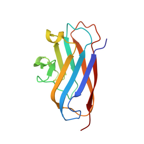 | |
UniProt | |||||
Find proteins for P39632 (Bacillus subtilis (strain 168)) Explore P39632 Go to UniProtKB: P39632 | |||||
Entity Groups | |||||
| Sequence Clusters | 30% Identity50% Identity70% Identity90% Identity95% Identity100% Identity | ||||
| UniProt Group | P39632 | ||||
Sequence AnnotationsExpand | |||||
| |||||
| Length ( Å ) | Angle ( ˚ ) |
|---|---|
| a = 121.405 | α = 90 |
| b = 128.306 | β = 90 |
| c = 84.327 | γ = 90 |
| Software Name | Purpose |
|---|---|
| MOLREP | phasing |
| REFMAC | refinement |
| PDB_EXTRACT | data extraction |
| MOLREP | phasing |
| xia2 | data reduction |
| xia2 | data scaling |
RCSB PDB (citation) is hosted by
RCSB PDB is a member of the

