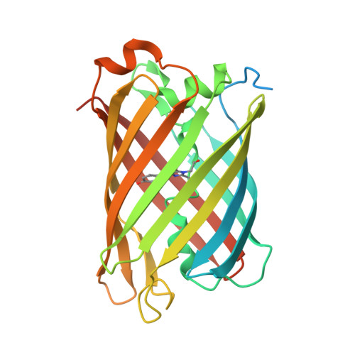
5LTP

Structure of the Yellow-Green Fluorescent Protein mNeonGreen from Branchiostoma lanceolatum at the acidic pH 4.5


Experimental Data Snapshot

  • Method: X-RAY DIFFRACTION
  • Resolution: 1.70 Å
  • R-Value Free: 0.211 
  • R-Value Work: 0.186 
  • R-Value Observed: 0.187 

wwPDB Validation   3D Report Full Report


This is version 1.1 of the entry. See complete history


Literature

Structural analysis of the bright monomeric yellow-green fluorescent protein mNeonGreen obtained by directed evolution.

Clavel, D.Gotthard, G.von Stetten, D.De Sanctis, D.Pasquier, H.Lambert, G.G.Shaner, N.C.Royant, A.

(2016) Acta Crystallogr D Struct Biol 72: 1298-1307

  • DOI: https://doi.org/10.1107/S2059798316018623
  • Primary Citation of Related Structures:  
    5LTP, 5LTQ, 5LTR

  • PubMed Abstract: 

    Until recently, genes coding for homologues of the autofluorescent protein GFP had only been identified in marine organisms from the phyla Cnidaria and Arthropoda. New fluorescent-protein genes have now been found in the phylum Chordata, coding for particularly bright oligomeric fluorescent proteins such as the tetrameric yellow fluorescent protein lanYFP from Branchiostoma lanceolatum. A successful monomerization attempt led to the development of the bright yellow-green fluorescent protein mNeonGreen. The structures of lanYFP and mNeonGreen have been determined and compared in order to rationalize the directed evolution process leading from a bright, tetrameric to a still bright, monomeric fluorescent protein. An unusual discolouration of crystals of mNeonGreen was observed after X-ray data collection, which was investigated using a combination of X-ray crystallography and UV-visible absorption and Raman spectroscopies, revealing the effects of specific radiation damage in the chromophore cavity. It is shown that X-rays rapidly lead to the protonation of the phenolate O atom of the chromophore and to the loss of its planarity at the methylene bridge.


  • Organizational Affiliation

    Université Grenoble Alpes, Institut de Biologie Structurale (IBS), F-38044 Grenoble, France.


Macromolecules
Find similar proteins by:  (by identity cutoff)  |  3D Structure
Entity ID: 1
MoleculeChains Sequence LengthOrganismDetailsImage
mNeonGreen
A, B, C, D, E
A, B, C, D, E, F
269Branchiostoma lanceolatumMutation(s): 0 
Gene Names: blFP-Y3
UniProt
Find proteins for A0A1S4NYF2 (Branchiostoma lanceolatum)
Explore A0A1S4NYF2 
Go to UniProtKB:  A0A1S4NYF2
Entity Groups  
Sequence Clusters30% Identity50% Identity70% Identity90% Identity95% Identity100% Identity
UniProt GroupA0A1S4NYF2
Sequence Annotations
Expand
  • Reference Sequence
Small Molecules
Modified Residues  1 Unique
IDChains TypeFormula2D DiagramParent
CR2
Query on CR2
A, B, C, D, E
A, B, C, D, E, F
L-PEPTIDE LINKINGC13 H13 N3 O4GLY, TYR, GLY
Experimental Data & Validation

Experimental Data

  • Method: X-RAY DIFFRACTION
  • Resolution: 1.70 Å
  • R-Value Free: 0.211 
  • R-Value Work: 0.186 
  • R-Value Observed: 0.187 
  • Space Group: P 21 21 21
Unit Cell:
Length ( Å )Angle ( ˚ )
a = 75.87α = 90
b = 127.6β = 90
c = 146.84γ = 90
Software Package:
Software NamePurpose
REFMACrefinement
XDSdata reduction
XSCALEdata scaling
PHASERphasing

Structure Validation

View Full Validation Report


Entry History 

Deposition Data

Revision History  (Full details and data files)

  • Version 1.0: 2016-12-21
    Type: Initial release
  • Version 1.1: 2024-01-17
    Changes: Data collection, Database references, Refinement description