5LSH

human lysozyme in complex with a tetrasaccharide fragment of the O-chain of LPS from Klebsiella pneumoniae


Experimental Data Snapshot

  • Method: X-RAY DIFFRACTION
  • Resolution: 1.06 Å
  • R-Value Free: 0.203 
  • R-Value Work: 0.180 
  • R-Value Observed: 0.181 

wwPDB Validation   3D Report Full Report


This is version 2.1 of the entry. See complete history


Literature

Lysozyme's lectin-like characteristics facilitates its immune defense function.

Zhang, R.Wu, L.Eckert, T.Burg-Roderfeld, M.Rojas-Macias, M.A.Lutteke, T.Krylov, V.B.Argunov, D.A.Datta, A.Markart, P.Guenther, A.Norden, B.Schauer, R.Bhunia, A.Enani, M.A.Billeter, M.Scheidig, A.J.Nifantiev, N.E.Siebert, H.C.

(2017) Q Rev Biophys 50: e9-e9

  • DOI: https://doi.org/10.1017/S0033583517000075
  • Primary Citation of Related Structures:  
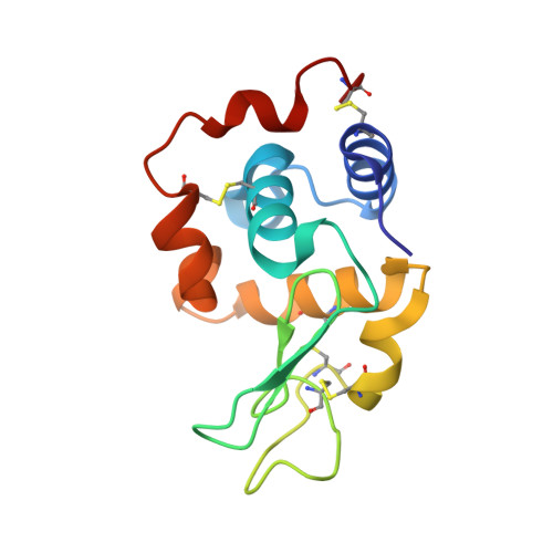
    5LSH

  • PubMed Abstract: 

    Interactions between human lysozyme (HL) and the lipopolysaccharide (LPS) of Klebsiella pneumoniae O1, a causative agent of lung infection, were identified by surface plasmon resonance. To characterize the molecular mechanism of this interaction, HL binding to synthetic disaccharides and tetrasaccharides representing one and two repeating units, respectively, of the O-chain of this LPS were studied. pH-dependent structural rearrangements of HL after interaction with the disaccharide were observed through nuclear magnetic resonance. The crystal structure of the HL-tetrasaccharide complex revealed carbohydrate chain packing into the A, B, C, and D binding sites of HL, which primarily occurred through residue-specific, direct or water-mediated hydrogen bonds and hydrophobic contacts. Overall, these results support a crucial role of the Glu35/Asp53/Trp63/Asp102 residues in HL binding to the tetrasaccharide. These observations suggest an unknown glycan-guided mechanism that underlies recognition of the bacterial cell wall by lysozyme and may complement the HL immune defense function.


  • Organizational Affiliation

    RI-B-NT Research Institute of Bioinformatics and Nanotechnology,Franziusallee 177, 24148 Kiel,Germany.


Macromolecules
Find similar proteins by:  (by identity cutoff)  |  3D Structure
Entity ID: 1
MoleculeChains Sequence LengthOrganismDetailsImage
Lysozyme C130Homo sapiensMutation(s): 0 
Gene Names: LYZLZM
EC: 3.2.1.17
UniProt & NIH Common Fund Data Resources
Find proteins for P61626 (Homo sapiens)
Explore P61626 
Go to UniProtKB:  P61626
PHAROS:  P61626
GTEx:  ENSG00000090382 
Entity Groups  
Sequence Clusters30% Identity50% Identity70% Identity90% Identity95% Identity100% Identity
UniProt GroupP61626
Sequence Annotations
Expand
  • Reference Sequence
Oligosaccharides

Help

Entity ID: 2
MoleculeChains Length2D Diagram Glycosylation3D Interactions
alpha-D-galactopyranose-(1-3)-beta-D-galactofuranose-(1-3)-alpha-D-galactopyranose-(1-3)-propyl beta-D-galactofuranoside
B
4N/AN/A
Glycosylation Resources
GlyTouCan:  G41009JY
GlyCosmos:  G41009JY
Experimental Data & Validation

Experimental Data

  • Method: X-RAY DIFFRACTION
  • Resolution: 1.06 Å
  • R-Value Free: 0.203 
  • R-Value Work: 0.180 
  • R-Value Observed: 0.181 
  • Space Group: P 21 21 21
Unit Cell:
Length ( Å )Angle ( ˚ )
a = 33.108α = 90
b = 56.04β = 90
c = 60.531γ = 90
Software Package:
Software NamePurpose
PHENIXrefinement
XDSdata reduction
XSCALEdata scaling
MOLREPphasing

Structure Validation

View Full Validation Report


Entry History 

Deposition Data

Revision History  (Full details and data files)

  • Version 1.0: 2017-06-21
    Type: Initial release
  • Version 1.1: 2017-12-27
    Changes: Database references
  • Version 2.0: 2020-07-29
    Type: Remediation
    Reason: Carbohydrate remediation
    Changes: Atomic model, Data collection, Derived calculations, Non-polymer description, Structure summary
  • Version 2.1: 2024-01-17
    Changes: Data collection, Database references, Refinement description, Structure summary