5KK2

Architecture of fully occupied GluA2 AMPA receptor - TARP complex elucidated by single particle cryo-electron microscopy


Experimental Data Snapshot

  • Method: ELECTRON MICROSCOPY
  • Resolution: 7.30 Å
  • Aggregation State: PARTICLE 
  • Reconstruction Method: SINGLE PARTICLE 

wwPDB Validation   3D Report Full Report


This is version 1.4 of the entry. See complete history


Literature

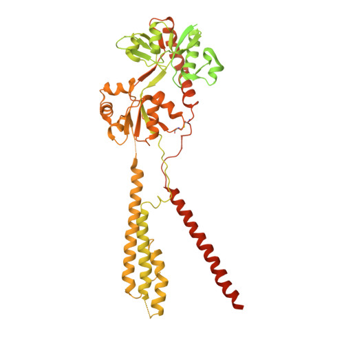
Architecture of fully occupied GluA2 AMPA receptor-TARP complex elucidated by cryo-EM.

Zhao, Y.Chen, S.Yoshioka, C.Baconguis, I.Gouaux, E.

(2016) Nature 536: 108-111

  • DOI: https://doi.org/10.1038/nature18961
  • Primary Citation of Related Structures:  
    5KK2

  • PubMed Abstract: 

    Fast excitatory neurotransmission in the mammalian central nervous system is largely carried out by AMPA-sensitive ionotropic glutamate receptors. Localized within the postsynaptic density of glutamatergic spines, AMPA receptors are composed of heterotetrameric receptor assemblies associated with auxiliary subunits, the most common of which are transmembrane AMPA receptor regulatory proteins (TARPs). The association of TARPs with AMPA receptors modulates receptor trafficking and the kinetics of receptor gating and pharmacology. Here we report the cryo-electron microscopy (cryo-EM) structure of the homomeric rat GluA2 AMPA receptor saturated with TARP γ2 subunits, which shows how the TARPs are arranged with four-fold symmetry around the ion channel domain and make extensive interactions with the M1, M2 and M4 transmembrane helices. Poised like partially opened ‘hands’ underneath the two-fold symmetric ligand-binding domain (LBD) 'clamshells', one pair of TARPs is juxtaposed near the LBD dimer interface, whereas the other pair is near the LBD dimer-dimer interface. The extracellular ‘domains’ of TARP are positioned to not only modulate LBD clamshell closure, but also affect conformational rearrangements of the LBD layer associated with receptor activation and desensitization, while the TARP transmembrane domains buttress the ion channel pore.


Macromolecules
Find similar proteins by:  (by identity cutoff)  |  3D Structure
Entity ID: 1
MoleculeChains Sequence LengthOrganismDetailsImage
Glutamate receptor 2
A, B, C, D
889Rattus norvegicusMutation(s): 1 
Gene Names: Gria2Glur2
Membrane Entity: Yes 
UniProt
Find proteins for P19491 (Rattus norvegicus)
Explore P19491 
Go to UniProtKB:  P19491
Entity Groups  
Sequence Clusters30% Identity50% Identity70% Identity90% Identity95% Identity100% Identity
UniProt GroupP19491
Sequence Annotations
Expand
  • Reference Sequence
Find similar proteins by:  (by identity cutoff)  |  3D Structure
Entity ID: 2
MoleculeChains Sequence LengthOrganismDetailsImage
Voltage-dependent calcium channel gamma-2 subunit
E, F, G, H
323Rattus norvegicusMutation(s): 0 
Gene Names: Cacng2
Membrane Entity: Yes 
UniProt
Find proteins for Q71RJ2 (Rattus norvegicus)
Explore Q71RJ2 
Go to UniProtKB:  Q71RJ2
Entity Groups  
Sequence Clusters30% Identity50% Identity70% Identity90% Identity95% Identity100% Identity
UniProt GroupQ71RJ2
Sequence Annotations
Expand
  • Reference Sequence
Experimental Data & Validation

Experimental Data

  • Method: ELECTRON MICROSCOPY
  • Resolution: 7.30 Å
  • Aggregation State: PARTICLE 
  • Reconstruction Method: SINGLE PARTICLE 
EM Software:
TaskSoftware PackageVersion
RECONSTRUCTIONRELION
MODEL REFINEMENTPHENIX

Structure Validation

View Full Validation Report


Entry History 

Deposition Data

Revision History  (Full details and data files)

  • Version 1.0: 2016-07-06
    Type: Initial release
  • Version 1.1: 2016-07-13
    Changes: Database references
  • Version 1.2: 2016-08-10
    Changes: Database references
  • Version 1.3: 2016-08-17
    Changes: Database references
  • Version 1.4: 2018-07-18
    Changes: Data collection