5JVL

C4-type pyruvate phospate dikinase: nucleotide binding domain with bound ATP analogue


Experimental Data Snapshot

  • Method: X-RAY DIFFRACTION
  • Resolution: 2.90 Å
  • R-Value Free: 0.232 
  • R-Value Work: 0.211 
  • R-Value Observed: 0.212 

wwPDB Validation   3D Report Full Report


Ligand Structure Quality Assessment 


This is version 1.3 of the entry. See complete history


Literature

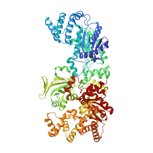
Structural intermediates and directionality of the swiveling motion of Pyruvate Phosphate Dikinase.

Minges, A.Ciupka, D.Winkler, C.Hoppner, A.Gohlke, H.Groth, G.

(2017) Sci Rep 7: 45389-45389

  • DOI: https://doi.org/10.1038/srep45389
  • Primary Citation of Related Structures:  
    5JVJ, 5JVL, 5JVN

  • PubMed Abstract: 

    Pyruvate phosphate dikinase (PPDK) is a vital enzyme in cellular energy metabolism catalyzing the ATP- and P i -dependent formation of phosphoenolpyruvate from pyruvate in C 4 -plants, but the reverse reaction forming ATP in bacteria and protozoa. The multi-domain enzyme is considered an efficient molecular machine that performs one of the largest single domain movements in proteins. However, a comprehensive understanding of the proposed swiveling domain motion has been limited by not knowing structural intermediates or molecular dynamics of the catalytic process. Here, we present crystal structures of PPDKs from Flaveria, a model genus for studying the evolution of C 4 -enzymes from phylogenetic ancestors. These structures resolve yet unknown conformational intermediates and provide the first detailed view on the large conformational transitions of the protein in the catalytic cycle. Independently performed unrestrained MD simulations and configurational free energy calculations also identified these intermediates. In all, our experimental and computational data reveal strict coupling of the CD swiveling motion to the conformational state of the NBD. Moreover, structural asymmetries and nucleotide binding states in the PPDK dimer support an alternate binding change mechanism for this intriguing bioenergetic enzyme.


  • Organizational Affiliation

    Cluster of Excellence on Plant Sciences (CEPLAS), Institute of Biochemical Plant Physiology, Heinrich Heine University Düsseldorf, 40204 Düsseldorf, Germany.


Macromolecules
Find similar proteins by:  (by identity cutoff)  |  3D Structure
Entity ID: 1
MoleculeChains Sequence LengthOrganismDetailsImage
Pyruvate, phosphate dikinase, chloroplastic
A, B, C, D
874Flaveria trinerviaMutation(s): 0 
Gene Names: PPDKPDK
EC: 2.7.9.1
UniProt
Find proteins for P22221 (Flaveria trinervia)
Explore P22221 
Go to UniProtKB:  P22221
Entity Groups  
Sequence Clusters30% Identity50% Identity70% Identity90% Identity95% Identity100% Identity
UniProt GroupP22221
Sequence Annotations
Expand
  • Reference Sequence
Small Molecules
Ligands 3 Unique
IDChains Name / Formula / InChI Key2D Diagram3D Interactions
6NQ
Query on 6NQ

Download Ideal Coordinates CCD File 
E [auth A],
K [auth C],
O [auth D]
2'-Bromo-2'-deoxyadenosine 5'-[beta,gamma-imide]triphosphoric acid
C10 H16 Br N6 O11 P3
GLCRVGHVCJLAHS-QYYRPYCUSA-N
PEP
Query on PEP

Download Ideal Coordinates CCD File 
G [auth A],
I [auth B],
M [auth C],
Q [auth D]
PHOSPHOENOLPYRUVATE
C3 H5 O6 P
DTBNBXWJWCWCIK-UHFFFAOYSA-N
MG
Query on MG

Download Ideal Coordinates CCD File 
F [auth A]
H [auth A]
J [auth B]
L [auth C]
N [auth C]
F [auth A],
H [auth A],
J [auth B],
L [auth C],
N [auth C],
P [auth D],
R [auth D]
MAGNESIUM ION
Mg
JLVVSXFLKOJNIY-UHFFFAOYSA-N
Experimental Data & Validation

Experimental Data

  • Method: X-RAY DIFFRACTION
  • Resolution: 2.90 Å
  • R-Value Free: 0.232 
  • R-Value Work: 0.211 
  • R-Value Observed: 0.212 
  • Space Group: P 1
  • Diffraction Data: https://doi.org/10.18430/m35jvl
Unit Cell:
Length ( Å )Angle ( ˚ )
a = 69.98α = 106.22
b = 108.45β = 101.81
c = 152.76γ = 98.32
Software Package:
Software NamePurpose
XDSdata scaling
XDSdata reduction
PHASERphasing
REFMACrefinement
PDB_EXTRACTdata extraction

Structure Validation

View Full Validation Report


Ligand Structure Quality Assessment 


Entry History 

Deposition Data

Revision History  (Full details and data files)

  • Version 1.0: 2017-04-05
    Type: Initial release
  • Version 1.1: 2017-04-12
    Changes: Database references
  • Version 1.2: 2020-04-22
    Changes: Database references
  • Version 1.3: 2024-01-10
    Changes: Data collection, Database references, Derived calculations, Refinement description