5DN6

ATP synthase from Paracoccus denitrificans


Experimental Data Snapshot

  • Method: X-RAY DIFFRACTION
  • Resolution: 3.98 Å
  • R-Value Free: 0.324 
  • R-Value Work: 0.295 
  • R-Value Observed: 0.297 

wwPDB Validation   3D Report Full Report


Ligand Structure Quality Assessment 


This is version 2.1 of the entry. See complete history


Literature

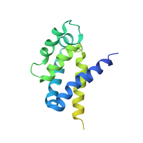
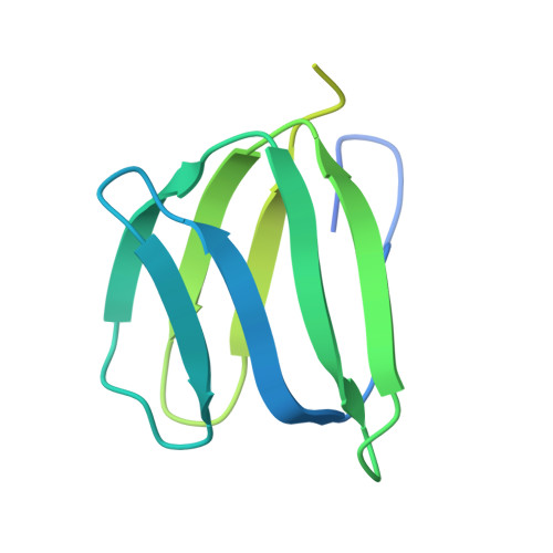
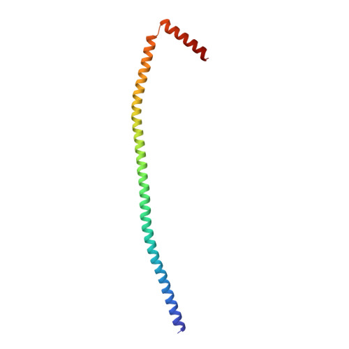
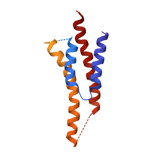
Structure of ATP synthase from Paracoccus denitrificans determined by X-ray crystallography at 4.0 angstrom resolution.

Morales-Rios, E.Montgomery, M.G.Leslie, A.G.Walker, J.E.

(2015) Proc Natl Acad Sci U S A 112: 13231-13236

  • DOI: https://doi.org/10.1073/pnas.1517542112
  • Primary Citation of Related Structures:  
    5DN6

  • PubMed Abstract: 

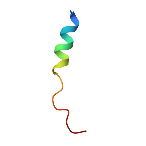
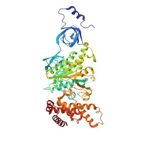
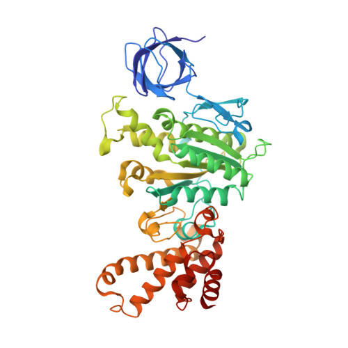
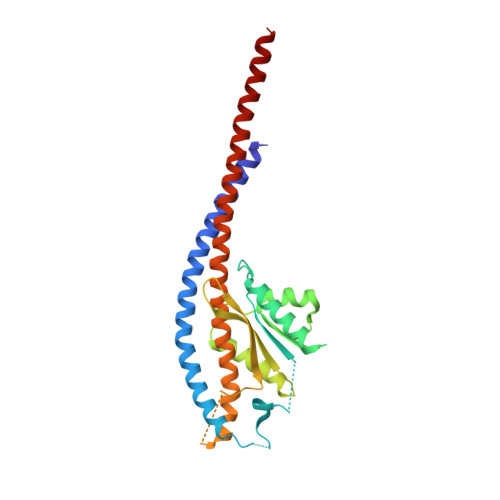
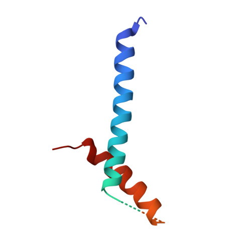
    The structure of the intact ATP synthase from the α-proteobacterium Paracoccus denitrificans, inhibited by its natural regulatory ζ-protein, has been solved by X-ray crystallography at 4.0 Å resolution. The ζ-protein is bound via its N-terminal α-helix in a catalytic interface in the F1 domain. The bacterial F1 domain is attached to the membrane domain by peripheral and central stalks. The δ-subunit component of the peripheral stalk binds to the N-terminal regions of two α-subunits. The stalk extends via two parallel long α-helices, one in each of the related b and b' subunits, down a noncatalytic interface of the F1 domain and interacts in an unspecified way with the a-subunit in the membrane domain. The a-subunit lies close to a ring of 12 c-subunits attached to the central stalk in the F1 domain, and, together, the central stalk and c-ring form the enzyme's rotor. Rotation is driven by the transmembrane proton-motive force, by a mechanism where protons pass through the interface between the a-subunit and c-ring via two half-channels in the a-subunit. These half-channels are probably located in a bundle of four α-helices in the a-subunit that are tilted at ∼30° to the plane of the membrane. Conserved polar residues in the two α-helices closest to the c-ring probably line the proton inlet path to an essential carboxyl group in the c-subunit in the proton uptake site and a proton exit path from the proton release site. The structure has provided deep insights into the workings of this extraordinary molecular machine.


  • Organizational Affiliation

    Medical Research Council Mitochondrial Biology Unit, Cambridge Biomedical Campus, Cambridge CB2 0XY, United Kingdom;


Macromolecules

Find similar proteins by:  Sequence   |   3D Structure  

Entity ID: 1
MoleculeChains Sequence LengthOrganismDetailsImage
Chain AA [auth 1]20Paracoccus denitrificansMutation(s): 0 
Sequence Annotations
Expand
  • Reference Sequence

Find similar proteins by:  Sequence   |   3D Structure  

Entity ID: 2
MoleculeChains Sequence LengthOrganismDetailsImage
Chain BB [auth 2]15Paracoccus denitrificansMutation(s): 0 
Sequence Annotations
Expand
  • Reference Sequence

Find similar proteins by:  Sequence   |   3D Structure  

Entity ID: 3
MoleculeChains Sequence LengthOrganismDetailsImage
Chain CC [auth 3]19Paracoccus denitrificansMutation(s): 0 
Sequence Annotations
Expand
  • Reference Sequence
Find similar proteins by:  (by identity cutoff)  |  3D Structure
Entity ID: 4
MoleculeChains Sequence LengthOrganismDetailsImage
ATP synthase subunit alphaD [auth A],
E [auth B],
F [auth C]
511Paracoccus denitrificansMutation(s): 0 
EC: 7.1.2.2
Membrane Entity: Yes 
UniProt
Find proteins for A1B8N8 (Paracoccus denitrificans (strain Pd 1222))
Explore A1B8N8 
Go to UniProtKB:  A1B8N8
Entity Groups  
Sequence Clusters30% Identity50% Identity70% Identity90% Identity95% Identity100% Identity
UniProt GroupA1B8N8
Sequence Annotations
Expand
  • Reference Sequence
Find similar proteins by:  (by identity cutoff)  |  3D Structure
Entity ID: 5
MoleculeChains Sequence LengthOrganismDetailsImage
ATP synthase subunit betaG [auth D],
H [auth E],
I [auth F]
474Paracoccus denitrificansMutation(s): 0 
EC: 7.1.2.2
Membrane Entity: Yes 
UniProt
Find proteins for A1B8P0 (Paracoccus denitrificans (strain Pd 1222))
Explore A1B8P0 
Go to UniProtKB:  A1B8P0
Entity Groups  
Sequence Clusters30% Identity50% Identity70% Identity90% Identity95% Identity100% Identity
UniProt GroupA1B8P0
Sequence Annotations
Expand
  • Reference Sequence
Find similar proteins by:  (by identity cutoff)  |  3D Structure
Entity ID: 6
MoleculeChains Sequence LengthOrganismDetailsImage
ATP synthase gamma chainJ [auth G]290Paracoccus denitrificansMutation(s): 0 
Membrane Entity: Yes 
UniProt
Find proteins for A1B8N9 (Paracoccus denitrificans (strain Pd 1222))
Explore A1B8N9 
Go to UniProtKB:  A1B8N9
Entity Groups  
Sequence Clusters30% Identity50% Identity70% Identity90% Identity95% Identity100% Identity
UniProt GroupA1B8N9
Sequence Annotations
Expand
  • Reference Sequence
Find similar proteins by:  (by identity cutoff)  |  3D Structure
Entity ID: 7
MoleculeChains Sequence LengthOrganismDetailsImage
ATP synthase subunit deltaK [auth H]188Paracoccus denitrificansMutation(s): 0 
Membrane Entity: Yes 
UniProt
Find proteins for A1B8N7 (Paracoccus denitrificans (strain Pd 1222))
Explore A1B8N7 
Go to UniProtKB:  A1B8N7
Entity Groups  
Sequence Clusters30% Identity50% Identity70% Identity90% Identity95% Identity100% Identity
UniProt GroupA1B8N7
Sequence Annotations
Expand
  • Reference Sequence
Find similar proteins by:  (by identity cutoff)  |  3D Structure
Entity ID: 8
MoleculeChains Sequence LengthOrganismDetailsImage
ATP synthase epsilon chainL [auth I]148Paracoccus denitrificansMutation(s): 0 
Membrane Entity: Yes 
UniProt
Find proteins for A1B8P1 (Paracoccus denitrificans (strain Pd 1222))
Explore A1B8P1 
Go to UniProtKB:  A1B8P1
Entity Groups  
Sequence Clusters30% Identity50% Identity70% Identity90% Identity95% Identity100% Identity
UniProt GroupA1B8P1
Sequence Annotations
Expand
  • Reference Sequence
Find similar proteins by:  (by identity cutoff)  |  3D Structure
Entity ID: 9
MoleculeChains Sequence LengthOrganismDetailsImage
ATP synthase F0 subcomplex C subunit77Paracoccus denitrificansMutation(s): 0 
Membrane Entity: Yes 
UniProt
Find proteins for A1B618 (Paracoccus denitrificans (strain Pd 1222))
Explore A1B618 
Go to UniProtKB:  A1B618
Entity Groups  
Sequence Clusters30% Identity50% Identity70% Identity90% Identity95% Identity100% Identity
UniProt GroupA1B618
Sequence Annotations
Expand
  • Reference Sequence
Find similar proteins by:  (by identity cutoff)  |  3D Structure
Entity ID: 10
MoleculeChains Sequence LengthOrganismDetailsImage
Chain VY [auth V]78Paracoccus denitrificansMutation(s): 0 
Sequence Annotations
Expand
  • Reference Sequence
Find similar proteins by:  (by identity cutoff)  |  3D Structure
Entity ID: 11
MoleculeChains Sequence LengthOrganismDetailsImage
Chain WZ [auth W]124Paracoccus denitrificansMutation(s): 0 
Sequence Annotations
Expand
  • Reference Sequence
Find similar proteins by:  (by identity cutoff)  |  3D Structure
Entity ID: 12
MoleculeChains Sequence LengthOrganismDetailsImage
ATP synthase subunit a,ATP synthase subunit a,ATP synthase subunit a,ATP synthase subunit aAA [auth X]283Paracoccus denitrificansMutation(s): 0 
Membrane Entity: Yes 
UniProt
Find proteins for A1B619 (Paracoccus denitrificans (strain Pd 1222))
Explore A1B619 
Go to UniProtKB:  A1B619
Entity Groups  
Sequence Clusters30% Identity50% Identity70% Identity90% Identity95% Identity100% Identity
UniProt GroupA1B619
Sequence Annotations
Expand
  • Reference Sequence
Find similar proteins by:  (by identity cutoff)  |  3D Structure
Entity ID: 13
MoleculeChains Sequence LengthOrganismDetailsImage
Chain YBA [auth Y]54Paracoccus denitrificansMutation(s): 0 
Sequence Annotations
Expand
  • Reference Sequence
Find similar proteins by:  (by identity cutoff)  |  3D Structure
Entity ID: 14
MoleculeChains Sequence LengthOrganismDetailsImage
Zeta inhibitor proteinCA [auth Z]104Paracoccus denitrificansMutation(s): 0 
UniProt
Find proteins for A1B602 (Paracoccus denitrificans (strain Pd 1222))
Explore A1B602 
Go to UniProtKB:  A1B602
Entity Groups  
Sequence Clusters30% Identity50% Identity70% Identity90% Identity95% Identity100% Identity
UniProt GroupA1B602
Sequence Annotations
Expand
  • Reference Sequence
Small Molecules
Ligands 3 Unique
IDChains Name / Formula / InChI Key2D Diagram3D Interactions
ATP
Query on ATP

Download Ideal Coordinates CCD File 
DA [auth A],
HA [auth C],
JA [auth D],
LA [auth F]
ADENOSINE-5'-TRIPHOSPHATE
C10 H16 N5 O13 P3
ZKHQWZAMYRWXGA-KQYNXXCUSA-N
ADP
Query on ADP

Download Ideal Coordinates CCD File 
FA [auth B]ADENOSINE-5'-DIPHOSPHATE
C10 H15 N5 O10 P2
XTWYTFMLZFPYCI-KQYNXXCUSA-N
MG
Query on MG

Download Ideal Coordinates CCD File 
EA [auth A],
GA [auth B],
IA [auth C],
KA [auth D],
MA [auth F]
MAGNESIUM ION
Mg
JLVVSXFLKOJNIY-UHFFFAOYSA-N
Experimental Data & Validation

Experimental Data

  • Method: X-RAY DIFFRACTION
  • Resolution: 3.98 Å
  • R-Value Free: 0.324 
  • R-Value Work: 0.295 
  • R-Value Observed: 0.297 
  • Space Group: P 1 21 1
Unit Cell:
Length ( Å )Angle ( ˚ )
a = 112.001α = 90
b = 187.943β = 97.44
c = 164.724γ = 90
Software Package:
Software NamePurpose
REFMACrefinement
Aimlessdata scaling
PHASERphasing

Structure Validation

View Full Validation Report


Ligand Structure Quality Assessment 


Entry History & Funding Information

Deposition Data


Funding OrganizationLocationGrant Number
Medical Research Council (United Kingdom)United KingdomMR/M009858/1
Medical Research Council (United Kingdom)United KingdomU105663150
Medical Research Council (United Kingdom)United KingdomU105184325

Revision History  (Full details and data files)

  • Version 1.0: 2015-10-28
    Type: Initial release
  • Version 1.1: 2017-04-05
    Changes: Other
  • Version 1.2: 2017-09-13
    Changes: Author supporting evidence
  • Version 1.3: 2018-01-17
    Changes: Experimental preparation
  • Version 2.0: 2018-07-04
    Changes: Advisory, Atomic model, Data collection, Database references, Derived calculations, Structure summary
  • Version 2.1: 2019-12-04
    Changes: Source and taxonomy, Structure summary