4JBU

1.65A structure of the T3SS tip protein LcrV (G28-D322, C273S) from Yersinia pestis


Experimental Data Snapshot

  • Method: X-RAY DIFFRACTION
  • Resolution: 1.65 Å
  • R-Value Free: 0.220 
  • R-Value Work: 0.177 
  • R-Value Observed: 0.179 

wwPDB Validation   3D Report Full Report


This is version 1.2 of the entry. See complete history


Literature

Structure of the Yersinia pestis tip protein LcrV refined to 1.65A resolution

Chaudhury, S.Battaile, K.P.Lovell, S.Plano, G.V.De Guzman, R.N.

(2013) Acta Crystallogr Sect F Struct Biol Cryst Commun 69: 477-481

  • DOI: https://doi.org/10.1107/S1744309113008579
  • Primary Citation of Related Structures:  
    4JBU

  • PubMed Abstract: 

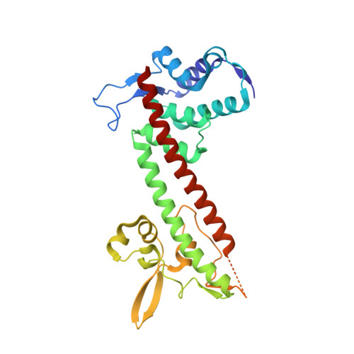
    The human pathogen Yersinia pestis requires the assembly of the type III secretion system (T3SS) for virulence. The structural component of the T3SS contains an external needle and a tip complex, which is formed by LcrV in Y. pestis. The structure of an LcrV triple mutant (K40A/D41A/K42A) in a C273S background has previously been reported to 2.2 Å resolution. Here, the crystal structure of LcrV without the triple mutation in a C273S background is reported at a higher resolution of 1.65 Å. Overall the two structures are similar, but there are also notable differences, particularly near the site of the triple mutation. The refined structure revealed a slight shift in the backbone positions of residues Gly28-Asn43 and displayed electron density in the loop region consisting of residues Ile46-Val63, which was disordered in the original structure. In addition, the helical turn region spanning residues Tyr77-Gln95 adopts a different orientation.


  • Organizational Affiliation

    Department of Molecular Biosciences, University of Kansas, Lawrence, KS 66045, USA.


Macromolecules
Find similar proteins by:  (by identity cutoff)  |  3D Structure
Entity ID: 1
MoleculeChains Sequence LengthOrganismDetailsImage
Virulence-associated V antigen298Yersinia pestisMutation(s): 1 
UniProt
Find proteins for P0C7U7 (Yersinia pestis)
Explore P0C7U7 
Go to UniProtKB:  P0C7U7
Entity Groups  
Sequence Clusters30% Identity50% Identity70% Identity90% Identity95% Identity100% Identity
UniProt GroupP0C7U7
Sequence Annotations
Expand
  • Reference Sequence
Small Molecules
Ligands 1 Unique
IDChains Name / Formula / InChI Key2D Diagram3D Interactions
PG4
Query on PG4

Download Ideal Coordinates CCD File 
B [auth A]TETRAETHYLENE GLYCOL
C8 H18 O5
UWHCKJMYHZGTIT-UHFFFAOYSA-N
Experimental Data & Validation

Experimental Data

  • Method: X-RAY DIFFRACTION
  • Resolution: 1.65 Å
  • R-Value Free: 0.220 
  • R-Value Work: 0.177 
  • R-Value Observed: 0.179 
  • Space Group: P 1 21 1
Unit Cell:
Length ( Å )Angle ( ˚ )
a = 39.553α = 90
b = 51.442β = 92
c = 65.178γ = 90
Software Package:
Software NamePurpose
SCALAdata scaling
PHASERphasing
PHENIXrefinement
PDB_EXTRACTdata extraction
XSCALEdata scaling

Structure Validation

View Full Validation Report


Entry History 

Deposition Data

Revision History  (Full details and data files)

  • Version 1.0: 2013-05-08
    Type: Initial release
  • Version 1.1: 2013-07-10
    Changes: Database references
  • Version 1.2: 2023-09-20
    Changes: Data collection, Database references, Derived calculations, Refinement description