4S1B

Crystal Structure of L. monocytogenes phosphodiesterase PgpH HD domain in complex with Cyclic-di-AMP


Experimental Data Snapshot

  • Method: X-RAY DIFFRACTION
  • Resolution: 2.10 Å
  • R-Value Free: 0.202 
  • R-Value Work: 0.163 
  • R-Value Observed: 0.165 

wwPDB Validation   3D Report Full Report


Ligand Structure Quality Assessment 


This is version 1.3 of the entry. See complete history


Literature

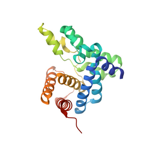
An HD-domain phosphodiesterase mediates cooperative hydrolysis of c-di-AMP to affect bacterial growth and virulence.

Huynh, T.N.Luo, S.Pensinger, D.Sauer, J.D.Tong, L.Woodward, J.J.

(2015) Proc Natl Acad Sci U S A 112: E747-E756

  • DOI: https://doi.org/10.1073/pnas.1416485112
  • Primary Citation of Related Structures:  
    4S1B, 4S1C

  • PubMed Abstract: 

    The nucleotide cyclic di-3',5'- adenosine monophosphate (c-di-AMP) was recently identified as an essential and widespread second messenger in bacterial signaling. Among c-di-AMP-producing bacteria, altered nucleotide levels result in several physiological defects and attenuated virulence. Thus, a detailed molecular understanding of c-di-AMP metabolism is of both fundamental and practical interest. Currently, c-di-AMP degradation is recognized solely among DHH-DHHA1 domain-containing phosphodiesterases. Using chemical proteomics, we identified the Listeria monocytogenes protein PgpH as a molecular target of c-di-AMP. Biochemical and structural studies revealed that the PgpH His-Asp (HD) domain bound c-di-AMP with high affinity and specifically hydrolyzed this nucleotide to 5'-pApA. PgpH hydrolysis activity was inhibited by ppGpp, indicating a cross-talk between c-di-AMP signaling and the stringent response. Genetic analyses supported coordinated regulation of c-di-AMP levels in and out of the host. Intriguingly, a L. monocytogenes mutant that lacks c-di-AMP phosphodiesterases exhibited elevated c-di-AMP levels, hyperinduced a host type-I IFN response, and was significantly attenuated for infection. Furthermore, PgpH homologs, which belong to the 7TMR-HD family, are widespread among hundreds of c-di-AMP synthesizing microorganisms. Thus, PgpH represents a broadly conserved class of c-di-AMP phosphodiesterase with possibly other physiological functions in this crucial signaling network.


  • Organizational Affiliation

    Department of Microbiology, University of Washington, Seattle, WA 98195;


Macromolecules
Find similar proteins by:  (by identity cutoff)  |  3D Structure
Entity ID: 1
MoleculeChains Sequence LengthOrganismDetailsImage
Lmo1466 proteinA,
B [auth D]
222Listeria monocytogenes EGD-eMutation(s): 0 
Gene Names: lmo1466pgpH
UniProt
Find proteins for Q8Y746 (Listeria monocytogenes serovar 1/2a (strain ATCC BAA-679 / EGD-e))
Explore Q8Y746 
Go to UniProtKB:  Q8Y746
Entity Groups  
Sequence Clusters30% Identity50% Identity70% Identity90% Identity95% Identity100% Identity
UniProt GroupQ8Y746
Sequence Annotations
Expand
  • Reference Sequence
Small Molecules
Binding Affinity Annotations 
IDSourceBinding Affinity
2BA Binding MOAD:  4S1B Kd: 300 (nM) from 1 assay(s)
Experimental Data & Validation

Experimental Data

  • Method: X-RAY DIFFRACTION
  • Resolution: 2.10 Å
  • R-Value Free: 0.202 
  • R-Value Work: 0.163 
  • R-Value Observed: 0.165 
  • Space Group: P 61
Unit Cell:
Length ( Å )Angle ( ˚ )
a = 124.286α = 90
b = 124.286β = 90
c = 58.019γ = 120
Software Package:
Software NamePurpose
DENZOdata reduction
SCALEPACKdata scaling
PHENIXrefinement
PDB_EXTRACTdata extraction
CBASSdata collection
HKL-2000data reduction
HKL-2000data scaling
SHARPphasing

Structure Validation

View Full Validation Report


Ligand Structure Quality Assessment 


Entry History 

Deposition Data

Revision History  (Full details and data files)

  • Version 1.0: 2015-02-11
    Type: Initial release
  • Version 1.1: 2015-03-04
    Changes: Database references
  • Version 1.2: 2017-11-22
    Changes: Refinement description
  • Version 1.3: 2024-02-28
    Changes: Data collection, Database references, Derived calculations, Structure summary