4QI2

X-ray structure of the ROQ domain from murine Roquin-1 in complex with a 23-mer Tnf-CDE RNA


Experimental Data Snapshot

  • Method: X-RAY DIFFRACTION
  • Resolution: 3.00 Å
  • R-Value Free: 0.247 
  • R-Value Work: 0.203 
  • R-Value Observed: 0.205 

wwPDB Validation   3D Report Full Report


This is version 1.2 of the entry. See complete history


Literature

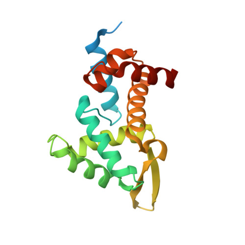
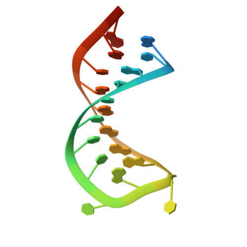
Structural basis for RNA recognition in roquin-mediated post-transcriptional gene regulation.

Schlundt, A.Heinz, G.A.Janowski, R.Geerlof, A.Stehle, R.Heissmeyer, V.Niessing, D.Sattler, M.

(2014) Nat Struct Mol Biol 21: 671-678

  • DOI: https://doi.org/10.1038/nsmb.2855
  • Primary Citation of Related Structures:  
    4QI0, 4QI2

  • PubMed Abstract: 

    Roquin function in T cells is essential for the prevention of autoimmune disease. Roquin interacts with the 3' untranslated regions (UTRs) of co-stimulatory receptors and controls T-cell activation and differentiation. Here we show that the N-terminal ROQ domain from mouse roquin adopts an extended winged-helix (WH) fold, which is sufficient for binding to the constitutive decay element (CDE) in the Tnf 3' UTR. The crystal structure of the ROQ domain in complex with a prototypical CDE RNA stem-loop reveals tight recognition of the RNA stem and its triloop. Surprisingly, roquin uses mainly non-sequence-specific contacts to the RNA, thus suggesting a relaxed CDE consensus and implicating a broader spectrum of target mRNAs than previously anticipated. Consistently with this, NMR and binding experiments with CDE-like stem-loops together with cell-based assays confirm roquin-dependent regulation of relaxed CDE consensus motifs in natural 3' UTRs.


  • Organizational Affiliation

    1] Biomolecular NMR Spectroscopy, Institute of Structural Biology, Helmholtz Zentrum München, Neuherberg, Germany. [2] Center for Integrated Protein Science Munich at Biomolecular NMR Spectroscopy, Department Chemie, Technische Universität München, Garching, Germany. [3].


Macromolecules

Find similar proteins by:  (by identity cutoff)  |  3D Structure
Entity ID: 1
MoleculeChains Sequence LengthOrganismDetailsImage
Roquin-1
A, B, C, D
180Mus musculusMutation(s): 0 
Gene Names: Gm551Kiaa2025Rc3h1
UniProt
Find proteins for Q4VGL6 (Mus musculus)
Explore Q4VGL6 
Go to UniProtKB:  Q4VGL6
Entity Groups  
Sequence Clusters30% Identity50% Identity70% Identity90% Identity95% Identity100% Identity
UniProt GroupQ4VGL6
Sequence Annotations
Expand
  • Reference Sequence

Find similar nucleic acids by:  Sequence   |   3D Structure  

Entity ID: 2
MoleculeChains LengthOrganismImage
RNA (5'-R(*AP*CP*AP*UP*GP*UP*UP*UP*UP*CP*UP*GP*UP*GP*AP*AP*AP*AP*CP*GP*GP*AP*G)-3')
E, F, G, H
23N/A
Sequence Annotations
Expand
  • Reference Sequence
Experimental Data & Validation

Experimental Data

  • Method: X-RAY DIFFRACTION
  • Resolution: 3.00 Å
  • R-Value Free: 0.247 
  • R-Value Work: 0.203 
  • R-Value Observed: 0.205 
  • Space Group: P 1
Unit Cell:
Length ( Å )Angle ( ˚ )
a = 56.56α = 105.68
b = 60.41β = 101.36
c = 84.37γ = 95.72
Software Package:
Software NamePurpose
DNAdata collection
PHASERphasing
REFMACrefinement
XDSdata reduction
SCALAdata scaling

Structure Validation

View Full Validation Report


Entry History 

Deposition Data

Revision History  (Full details and data files)

  • Version 1.0: 2014-07-16
    Type: Initial release
  • Version 1.1: 2014-08-20
    Changes: Database references
  • Version 1.2: 2023-09-20
    Changes: Data collection, Database references, Refinement description