4OUR

Crystal structure of Arabidopsis thaliana phytochrome B photosensory module


Experimental Data Snapshot

  • Method: X-RAY DIFFRACTION
  • Resolution: 3.40 Å
  • R-Value Free: 0.270 
  • R-Value Work: 0.242 
  • R-Value Observed: 0.244 

wwPDB Validation   3D Report Full Report


Ligand Structure Quality Assessment 


This is version 2.1 of the entry. See complete history


Literature

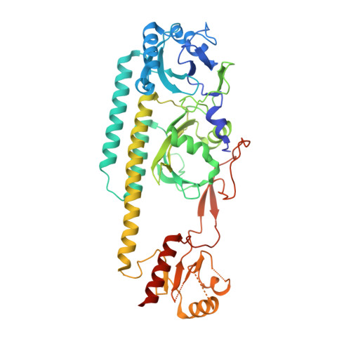
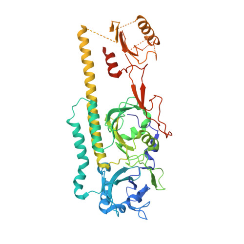
Crystal structure of the photosensing module from a red/far-red light-absorbing plant phytochrome.

Burgie, E.S.Bussell, A.N.Walker, J.M.Dubiel, K.Vierstra, R.D.

(2014) Proc Natl Acad Sci U S A 111: 10179-10184

  • DOI: https://doi.org/10.1073/pnas.1403096111
  • Primary Citation of Related Structures:  
    4OUR

  • PubMed Abstract: 

    Many aspects of plant photomorphogenesis are controlled by the phytochrome (Phy) family of bilin-containing photoreceptors that detect red and far-red light by photointerconversion between a dark-adapted Pr state and a photoactivated Pfr state. Whereas 3D models of prokaryotic Phys are available, models of their plant counterparts have remained elusive. Here, we present the crystal structure of the photosensing module (PSM) from a seed plant Phy in the Pr state using the PhyB isoform from Arabidopsis thaliana. The PhyB PSM crystallized as a head-to-head dimer with strong structural homology to its bacterial relatives, including a 5(Z)syn, 10(Z)syn, 15(Z)anti configuration of the phytochromobilin chromophore buried within the cGMP phosphodiesterase/adenylyl cyclase/FhlA (GAF) domain, and a well-ordered hairpin protruding from the Phy-specific domain toward the bilin pocket. However, its Per/Arnt/Sim (PAS) domain, knot region, and helical spine show distinct structural differences potentially important to signaling. Included is an elongated helical spine, an extended β-sheet connecting the GAF domain and hairpin stem, and unique interactions between the region upstream of the PAS domain knot and the bilin A and B pyrrole rings. Comparisons of this structure with those from bacterial Phys combined with mutagenic studies support a toggle model for photoconversion that engages multiple features within the PSM to stabilize the Pr and Pfr end states after rotation of the D pyrrole ring. Taken together, this Arabidopsis PhyB structure should enable molecular insights into plant Phy signaling and provide an essential scaffold to redesign their activities for agricultural benefit and as optogenetic reagents.


  • Organizational Affiliation

    Department of Genetics, University of Wisconsin-Madison, Madison, WI 53706.


Macromolecules
Find similar proteins by:  (by identity cutoff)  |  3D Structure
Entity ID: 1
MoleculeChains Sequence LengthOrganismDetailsImage
Phytochrome B544Arabidopsis thalianaMutation(s): 0 
Gene Names: At2g18790HY3MSF3.17PHYB
UniProt
Find proteins for P14713 (Arabidopsis thaliana)
Explore P14713 
Go to UniProtKB:  P14713
Entity Groups  
Sequence Clusters30% Identity50% Identity70% Identity90% Identity95% Identity100% Identity
UniProt GroupP14713
Sequence Annotations
Expand
  • Reference Sequence
Find similar proteins by:  (by identity cutoff)  |  3D Structure
Entity ID: 2
MoleculeChains Sequence LengthOrganismDetailsImage
Phytochrome B544Arabidopsis thalianaMutation(s): 0 
Gene Names: PHYBHY3At2g18790MSF3.17
UniProt
Find proteins for P14713 (Arabidopsis thaliana)
Explore P14713 
Go to UniProtKB:  P14713
Entity Groups  
Sequence Clusters30% Identity50% Identity70% Identity90% Identity95% Identity100% Identity
UniProt GroupP14713
Sequence Annotations
Expand
  • Reference Sequence
Small Molecules
Ligands 4 Unique
IDChains Name / Formula / InChI Key2D Diagram3D Interactions
O6E
Query on O6E

Download Ideal Coordinates CCD File 
K [auth A],
R [auth B]
3-[5-[[(3~{R},4~{R})-3-ethyl-4-methyl-5-oxidanylidene-3,4-dihydropyrrol-2-yl]methyl]-2-[[5-[(4-ethyl-3-methyl-5-oxidanylidene-pyrrol-2-yl)methyl]-3-(3-hydroxy-3-oxopropyl)-4-methyl-1~{H}-pyrrol-2-yl]methyl]-4-methyl-1~{H}-pyrrol-3-yl]propanoic acid
C33 H38 N4 O6
SRFSSETUPHUBBC-GMXXPEQVSA-N
PGE
Query on PGE

Download Ideal Coordinates CCD File 
Q [auth B]TRIETHYLENE GLYCOL
C6 H14 O4
ZIBGPFATKBEMQZ-UHFFFAOYSA-N
SO4
Query on SO4

Download Ideal Coordinates CCD File 
C [auth A]
D [auth A]
E [auth A]
F [auth A]
G [auth A]
C [auth A],
D [auth A],
E [auth A],
F [auth A],
G [auth A],
H [auth A],
I [auth A],
M [auth B],
N [auth B],
O [auth B],
P [auth B]
SULFATE ION
O4 S
QAOWNCQODCNURD-UHFFFAOYSA-L
GOL
Query on GOL

Download Ideal Coordinates CCD File 
J [auth A],
L [auth B]
GLYCEROL
C3 H8 O3
PEDCQBHIVMGVHV-UHFFFAOYSA-N
Experimental Data & Validation

Experimental Data

  • Method: X-RAY DIFFRACTION
  • Resolution: 3.40 Å
  • R-Value Free: 0.270 
  • R-Value Work: 0.242 
  • R-Value Observed: 0.244 
  • Space Group: P 41 21 2
Unit Cell:
Length ( Å )Angle ( ˚ )
a = 127.502α = 90
b = 127.502β = 90
c = 300.772γ = 90
Software Package:
Software NamePurpose
HKL-2000data collection
PHASERphasing
PHENIXrefinement
HKL-2000data reduction
HKL-2000data scaling

Structure Validation

View Full Validation Report


Ligand Structure Quality Assessment 


Entry History 

Deposition Data

Revision History  (Full details and data files)

  • Version 1.0: 2014-07-16
    Type: Initial release
  • Version 1.1: 2014-07-30
    Changes: Database references
  • Version 2.0: 2021-10-13
    Changes: Advisory, Atomic model, Data collection, Database references, Derived calculations, Non-polymer description, Structure summary
  • Version 2.1: 2023-09-20
    Changes: Data collection, Refinement description