4K95

Crystal Structure of Parkin


Experimental Data Snapshot

  • Method: X-RAY DIFFRACTION
  • Resolution: 6.50 Å
  • R-Value Free: 0.327 
  • R-Value Work: 0.307 
  • R-Value Observed: 0.308 

wwPDB Validation   3D Report Full Report


This is version 1.3 of the entry. See complete history


Literature

Structure of parkin reveals mechanisms for ubiquitin ligase activation.

Trempe, J.F.Sauve, V.Grenier, K.Seirafi, M.Tang, M.Y.Menade, M.Al-Abdul-Wahid, S.Krett, J.Wong, K.Kozlov, G.Nagar, B.Fon, E.A.Gehring, K.

(2013) Science 340: 1451-1455

  • DOI: https://doi.org/10.1126/science.1237908
  • Primary Citation of Related Structures:  
    4K7D, 4K95

  • PubMed Abstract: 

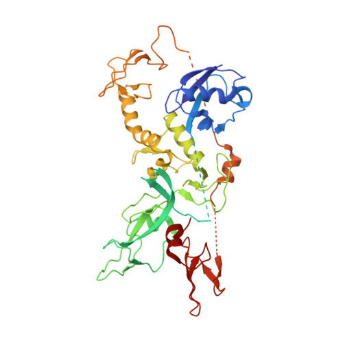
    Mutations in the PARK2 (parkin) gene are responsible for an autosomal recessive form of Parkinson's disease. The parkin protein is a RING-in-between-RING E3 ubiquitin ligase that exhibits low basal activity. We describe the crystal structure of full-length rat parkin. The structure shows parkin in an autoinhibited state and provides insight into how it is activated. RING0 occludes the ubiquitin acceptor site Cys(431) in RING2, whereas a repressor element of parkin binds RING1 and blocks its E2-binding site. Mutations that disrupted these inhibitory interactions activated parkin both in vitro and in cells. Parkin is neuroprotective, and these findings may provide a structural and mechanistic framework for enhancing parkin activity.


  • Organizational Affiliation

    McGill Parkinson Program, Department of Neurology and Neurosurgery, Montreal Neurological Institute, McGill University, Montréal, Québec, Canada.


Macromolecules
Find similar proteins by:  (by identity cutoff)  |  3D Structure
Entity ID: 1
MoleculeChains Sequence LengthOrganismDetailsImage
E3 ubiquitin-protein ligase parkin
A, B, C, D, E
A, B, C, D, E, F, G, H, I, J, K, L
470Rattus norvegicusMutation(s): 0 
Gene Names: Park2Prkn
EC: 6.3.2
UniProt
Find proteins for Q9JK66 (Rattus norvegicus)
Explore Q9JK66 
Go to UniProtKB:  Q9JK66
Entity Groups  
Sequence Clusters30% Identity50% Identity70% Identity90% Identity95% Identity100% Identity
UniProt GroupQ9JK66
Sequence Annotations
Expand
  • Reference Sequence
Small Molecules
Ligands 1 Unique
IDChains Name / Formula / InChI Key2D Diagram3D Interactions
ZN
Query on ZN

Download Ideal Coordinates CCD File 
AA [auth B]
AB [auth F]
AC [auth I]
AD [auth L]
BA [auth B]
AA [auth B],
AB [auth F],
AC [auth I],
AD [auth L],
BA [auth B],
BB [auth F],
BC [auth I],
BD [auth L],
CA [auth C],
CB [auth F],
CC [auth I],
CD [auth L],
DA [auth C],
DB [auth F],
DC [auth I],
DD [auth L],
EA [auth C],
EB [auth F],
EC [auth I],
FA [auth C],
FB [auth F],
FC [auth I],
GA [auth C],
GB [auth F],
GC [auth J],
HA [auth C],
HB [auth F],
HC [auth J],
IA [auth C],
IB [auth G],
IC [auth J],
JA [auth C],
JB [auth G],
JC [auth J],
KA [auth D],
KB [auth G],
KC [auth J],
LA [auth D],
LB [auth G],
LC [auth J],
M [auth A],
MA [auth D],
MB [auth G],
MC [auth J],
N [auth A],
NA [auth D],
NB [auth G],
NC [auth J],
O [auth A],
OA [auth D],
OB [auth G],
OC [auth K],
P [auth A],
PA [auth D],
PB [auth G],
PC [auth K],
Q [auth A],
QA [auth D],
QB [auth H],
QC [auth K],
R [auth A],
RA [auth D],
RB [auth H],
RC [auth K],
S [auth A],
SA [auth E],
SB [auth H],
SC [auth K],
T [auth A],
TA [auth E],
TB [auth H],
TC [auth K],
U [auth B],
UA [auth E],
UB [auth H],
UC [auth K],
V [auth B],
VA [auth E],
VB [auth H],
VC [auth K],
W [auth B],
WA [auth E],
WB [auth H],
WC [auth L],
X [auth B],
XA [auth E],
XB [auth H],
XC [auth L],
Y [auth B],
YA [auth E],
YB [auth I],
YC [auth L],
Z [auth B],
ZA [auth E],
ZB [auth I],
ZC [auth L]
ZINC ION
Zn
PTFCDOFLOPIGGS-UHFFFAOYSA-N
Experimental Data & Validation

Experimental Data

  • Method: X-RAY DIFFRACTION
  • Resolution: 6.50 Å
  • R-Value Free: 0.327 
  • R-Value Work: 0.307 
  • R-Value Observed: 0.308 
  • Space Group: P 21 21 2
Unit Cell:
Length ( Å )Angle ( ˚ )
a = 208.598α = 90
b = 277.439β = 90
c = 125.891γ = 90
Software Package:
Software NamePurpose
DENZOdata reduction
SCALEPACKdata scaling
PHENIXrefinement
PDB_EXTRACTdata extraction
ADSCdata collection
HKL-2000data reduction
HKL-2000data scaling
PHASERphasing

Structure Validation

View Full Validation Report


Entry History 

Deposition Data

Revision History  (Full details and data files)

  • Version 1.0: 2013-05-15
    Type: Initial release
  • Version 1.1: 2013-05-22
    Changes: Database references
  • Version 1.2: 2013-07-10
    Changes: Database references
  • Version 1.3: 2023-09-20
    Changes: Data collection, Database references, Derived calculations, Refinement description