Structure and regulon of Campylobacter jejuni ferric uptake regulator Fur define apo-Fur regulation.
Butcher, J., Sarvan, S., Brunzelle, J.S., Couture, J.F., Stintzi, A.(2012) Proc Natl Acad Sci U S A 109: 10047-10052
- PubMed: 22665794
- DOI: https://doi.org/10.1073/pnas.1118321109
- Primary Citation of Related Structures:
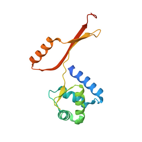
4ETS - PubMed Abstract:
The full regulatory potential of the ferric uptake regulator (Fur) family of proteins remains undefined despite over 20 years of study. We report herein an integrated approach that combines both genome-wide technologies and structural studies to define the role of Fur in Campylobacter jejuni (Cj). CjFur ChIP-chip assays identified 95 genomic loci bound by CjFur associated with functions as diverse as iron acquisition, flagellar biogenesis, and non-iron ion transport. Comparative analysis with transcriptomic data revealed that CjFur regulation extends beyond solely repression and also includes both gene activation and iron-independent regulation. Computational analysis revealed the presence of an elongated holo-Fur repression motif along with a divergent holo-Fur activation motif. This diversity of CjFur DNA-binding elements is supported by the crystal structure of CjFur, which revealed a unique conformation of its DNA-binding domain and the absence of metal in the regulatory site. Strikingly, our results indicate that the apo-CjFur structure retains the canonical V-shaped dimer reminiscent of previously characterized holo-Fur proteins enabling DNA interaction. This conformation stems from a structurally unique hinge domain that is poised to further contribute to CjFur's regulatory functions by modulating the orientation of the DNA-binding domain upon binding of iron. The unique features of the CjFur crystal structure rationalize the binding sequence diversity that was uncovered during ChIP-chip analysis and defines apo-Fur regulation.
Organizational Affiliation:
Department of Biochemistry, Microbiology and Immunology, Ottawa Institute of Systems Biology, University of Ottawa, Ottawa, ON, Canada, K1H 8M5.