The structure of the oligomerization domain of Lsr2 from Mycobacterium tuberculosis reveals a mechanism for chromosome organization and protection.
Summers, E.L., Meindl, K., Uson, I., Mitra, A.K., Radjainia, M., Colangeli, R., Alland, D., Arcus, V.L.(2012) PLoS One 7: e38542-e38542
- PubMed: 22719899
- DOI: https://doi.org/10.1371/journal.pone.0038542
- Primary Citation of Related Structures:
4E1P, 4E1R - PubMed Abstract:
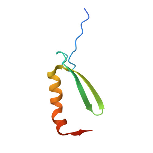
Lsr2 is a small DNA-binding protein present in mycobacteria and related actinobacteria that regulates gene expression and influences the organization of bacterial chromatin. Lsr2 is a dimer that binds to AT-rich regions of chromosomal DNA and physically protects DNA from damage by reactive oxygen intermediates (ROI). A recent structure of the C-terminal DNA-binding domain of Lsr2 provides a rationale for its interaction with the minor groove of DNA, its preference for AT-rich tracts, and its similarity to other bacterial nucleoid-associated DNA-binding domains. In contrast, the details of Lsr2 dimerization (and oligomerization) via its N-terminal domain, and the mechanism of Lsr2-mediated chromosomal cross-linking and protection is unknown. We have solved the structure of the N-terminal domain of Lsr2 (N-Lsr2) at 1.73 Å resolution using crystallographic ab initio approaches. The structure shows an intimate dimer of two ß-ß-a motifs with no close homologues in the structural databases. The organization of individual N-Lsr2 dimers in the crystal also reveals a mechanism for oligomerization. Proteolytic removal of three N-terminal residues from Lsr2 results in the formation of an anti-parallel β-sheet between neighboring molecules and the formation of linear chains of N-Lsr2. Oligomerization can be artificially induced using low concentrations of trypsin and the arrangement of N-Lsr2 into long chains is observed in both monoclinic and hexagonal crystallographic space groups. In solution, oligomerization of N-Lsr2 is also observed following treatment with trypsin. A change in chromosomal topology after the addition of trypsin to full-length Lsr2-DNA complexes and protection of DNA towards DNAse digestion can be observed using electron microscopy and electrophoresis. These results suggest a mechanism for oligomerization of Lsr2 via protease-activation leading to chromosome compaction and protection, and concomitant down-regulation of large numbers of genes. This mechanism is likely to be relevant under conditions of stress where cellular proteases are known to be upregulated.
Organizational Affiliation:
Department of Biological Sciences, University of Waikato, Hamilton, New Zealand.