4DOU

Crystal Structure of a Single-chain Trimer of Human Adiponectin Globular Domain


Experimental Data Snapshot

  • Method: X-RAY DIFFRACTION
  • Resolution: 2.00 Å
  • R-Value Free: 0.257 
  • R-Value Work: 0.200 
  • R-Value Observed: 0.203 

wwPDB Validation   3D Report Full Report


This is version 1.3 of the entry. See complete history


Literature

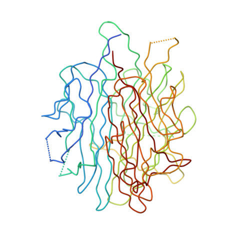
Crystal structure of a single-chain trimer of human adiponectin globular domain.

Min, X.Lemon, B.Tang, J.Liu, Q.Zhang, R.Walker, N.Li, Y.Wang, Z.

(2012) FEBS Lett 586: 912-917

  • DOI: https://doi.org/10.1016/j.febslet.2012.02.024
  • Primary Citation of Related Structures:  
    4DOU

  • PubMed Abstract: 

    Adiponectin is increasingly recognized as a potential therapeutic agent for the treatment of diabetes and other metabolic diseases. It circulates in plasma as homotrimers and higher-order oliogomers of homotrimers. To facilitate the production of active recombinant adiponectin as a therapeutic tool, we designed a single-chain globular domain adiponectin (sc-gAd) in which three monomer sequences are linked together in tandem to form one contiguous polypeptide. Here, we present the crystal structure of human sc-gAd at 2.0Å resolution. The structure reveals a similar trimeric topology to that of mouse gAd protein. Trimer formation is further rigidified by three calcium ions.


  • Organizational Affiliation

    Department of Molecular Structure, Amgen Inc., South San Francisco, CA 94080, USA.


Macromolecules
Find similar proteins by:  (by identity cutoff)  |  3D Structure
Entity ID: 1
MoleculeChains Sequence LengthOrganismDetailsImage
Adiponectin442Homo sapiensMutation(s): 0 
Gene Names: ADIPOQACDCACRP30APM1GBP28adiponectin
UniProt & NIH Common Fund Data Resources
Find proteins for Q15848 (Homo sapiens)
Explore Q15848 
Go to UniProtKB:  Q15848
PHAROS:  Q15848
GTEx:  ENSG00000181092 
Entity Groups  
Sequence Clusters30% Identity50% Identity70% Identity90% Identity95% Identity100% Identity
UniProt GroupQ15848
Sequence Annotations
Expand
  • Reference Sequence
Small Molecules
Ligands 4 Unique
IDChains Name / Formula / InChI Key2D Diagram3D Interactions
GOL
Query on GOL

Download Ideal Coordinates CCD File 
F [auth A]GLYCEROL
C3 H8 O3
PEDCQBHIVMGVHV-UHFFFAOYSA-N
EDO
Query on EDO

Download Ideal Coordinates CCD File 
G [auth A],
H [auth A],
I [auth A],
J [auth A],
K [auth A]
1,2-ETHANEDIOL
C2 H6 O2
LYCAIKOWRPUZTN-UHFFFAOYSA-N
CA
Query on CA

Download Ideal Coordinates CCD File 
B [auth A],
C [auth A],
D [auth A]
CALCIUM ION
Ca
BHPQYMZQTOCNFJ-UHFFFAOYSA-N
NA
Query on NA

Download Ideal Coordinates CCD File 
E [auth A]SODIUM ION
Na
FKNQFGJONOIPTF-UHFFFAOYSA-N
Experimental Data & Validation

Experimental Data

  • Method: X-RAY DIFFRACTION
  • Resolution: 2.00 Å
  • R-Value Free: 0.257 
  • R-Value Work: 0.200 
  • R-Value Observed: 0.203 
  • Space Group: P 1
Unit Cell:
Length ( Å )Angle ( ˚ )
a = 48.31α = 117.23
b = 49.63β = 101.68
c = 50.46γ = 105.31
Software Package:
Software NamePurpose
BOSdata collection
PHASERphasing
REFMACrefinement
MOSFLMdata reduction
SCALAdata scaling

Structure Validation

View Full Validation Report


Entry History 

Deposition Data

Revision History  (Full details and data files)

  • Version 1.0: 2012-02-29
    Type: Initial release
  • Version 1.1: 2012-05-02
    Changes: Database references
  • Version 1.2: 2017-05-31
    Changes: Structure summary
  • Version 1.3: 2023-09-13
    Changes: Data collection, Database references, Derived calculations, Refinement description