4CMQ

Crystal structure of Mn-bound S.pyogenes Cas9


Experimental Data Snapshot

  • Method: X-RAY DIFFRACTION
  • Resolution: 3.09 Å
  • R-Value Free: 0.277 
  • R-Value Work: 0.249 
  • R-Value Observed: 0.250 

wwPDB Validation   3D Report Full Report


This is version 1.3 of the entry. See complete history


Literature

Structures of Cas9 Endonucleases Reveal RNA- Mediated Conformational Activation

Jinek, M.Jiang, F.Taylor, D.W.Sternberg, S.H.Kaya, E.Ma, E.Anders, C.Hauer, M.Zhou, K.Lin, S.Kaplan, M.Iavarone, A.T.Charpentier, E.Nogales, E.Doudna, J.A.

(2014) Science 343: 47997

  • DOI: https://doi.org/10.1126/science.1247997
  • Primary Citation of Related Structures:  
    4CMP, 4CMQ, 4OGC, 4OGE

  • PubMed Abstract: 

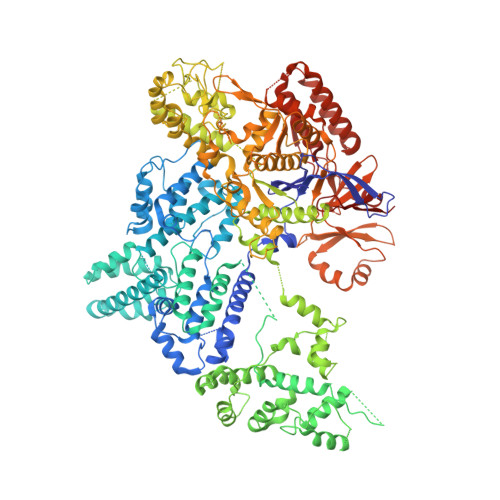
    Type II CRISPR (clustered regularly interspaced short palindromic repeats)-Cas (CRISPR-associated) systems use an RNA-guided DNA endonuclease, Cas9, to generate double-strand breaks in invasive DNA during an adaptive bacterial immune response. Cas9 has been harnessed as a powerful tool for genome editing and gene regulation in many eukaryotic organisms. We report 2.6 and 2.2 angstrom resolution crystal structures of two major Cas9 enzyme subtypes, revealing the structural core shared by all Cas9 family members. The architectures of Cas9 enzymes define nucleic acid binding clefts, and single-particle electron microscopy reconstructions show that the two structural lobes harboring these clefts undergo guide RNA-induced reorientation to form a central channel where DNA substrates are bound. The observation that extensive structural rearrangements occur before target DNA duplex binding implicates guide RNA loading as a key step in Cas9 activation.


  • Organizational Affiliation

    Department of Biochemistry, University of Zurich, CH-8057 Zurich, Switzerland.


Macromolecules
Find similar proteins by:  (by identity cutoff)  |  3D Structure
Entity ID: 1
MoleculeChains Sequence LengthOrganismDetailsImage
CRISPR-ASSOCIATED ENDONUCLEASE CAS9/CSN1
A, B
1,372Streptococcus pyogenes serotype M1Mutation(s): 0 
EC: 3.1
UniProt
Find proteins for Q99ZW2 (Streptococcus pyogenes serotype M1)
Explore Q99ZW2 
Go to UniProtKB:  Q99ZW2
Entity Groups  
Sequence Clusters30% Identity50% Identity70% Identity90% Identity95% Identity100% Identity
UniProt GroupQ99ZW2
Sequence Annotations
Expand
  • Reference Sequence
Small Molecules
Ligands 2 Unique
IDChains Name / Formula / InChI Key2D Diagram3D Interactions
SO4
Query on SO4

Download Ideal Coordinates CCD File 
J [auth A]
K [auth A]
L [auth A]
S [auth B]
T [auth B]
J [auth A],
K [auth A],
L [auth A],
S [auth B],
T [auth B],
U [auth B]
SULFATE ION
O4 S
QAOWNCQODCNURD-UHFFFAOYSA-L
MN
Query on MN

Download Ideal Coordinates CCD File 
C [auth A]
D [auth A]
E [auth A]
F [auth A]
G [auth A]
C [auth A],
D [auth A],
E [auth A],
F [auth A],
G [auth A],
H [auth A],
I [auth A],
M [auth B],
N [auth B],
O [auth B],
P [auth B],
Q [auth B],
R [auth B]
MANGANESE (II) ION
Mn
WAEMQWOKJMHJLA-UHFFFAOYSA-N
Experimental Data & Validation

Experimental Data

  • Method: X-RAY DIFFRACTION
  • Resolution: 3.09 Å
  • R-Value Free: 0.277 
  • R-Value Work: 0.249 
  • R-Value Observed: 0.250 
  • Space Group: P 21 21 2
Unit Cell:
Length ( Å )Angle ( ˚ )
a = 159.88α = 90
b = 210.12β = 90
c = 90.67γ = 90
Software Package:
Software NamePurpose
PHENIXrefinement
XDSdata reduction
XSCALEdata scaling
PHASERphasing

Structure Validation

View Full Validation Report


Entry History 

Revision History  (Full details and data files)

  • Version 1.0: 2014-02-12
    Type: Initial release
  • Version 1.1: 2014-02-19
    Changes: Database references
  • Version 1.2: 2014-03-26
    Changes: Database references
  • Version 1.3: 2023-12-20
    Changes: Data collection, Database references, Derived calculations, Other, Refinement description