Molecular Mechanism of Peroxisome Proliferator-Activated Receptor Alpha Activation by Wy14643: A New Mode of Ligand Recognition and Receptor Stabilization
Bernardes, A., T Souza, P.C., Muniz, J.R.C., Ricci, C.G., Ayers, S.D., Parekh, N.M., Godoy, A.S., Trivella, D.B.B., Reinach, P., Webb, P., Skaf, M.S., Polikarpov, I.(2013) J Mol Biol 425: 2878
- PubMed: 23707408
- DOI: https://doi.org/10.1016/j.jmb.2013.05.010
- Primary Citation of Related Structures:
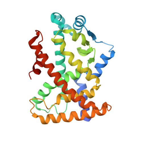
4BCR - PubMed Abstract:
Peroxisome proliferator-activated receptors (PPARs) are members of a superfamily of nuclear transcription factors. They are involved in mediating numerous physiological effects in humans, including glucose and lipid metabolism. PPARα ligands effectively treat dyslipidemia and have significant antiinflammatory and anti-atherosclerotic activities. These effects and their ligand-dependent activity make nuclear receptors obvious targets for drug design. Here, we present the structure of the human PPARα in complex with WY14643, a member of fibrate class of drug, and a widely used PPAR activator. The crystal structure of this complex suggests that WY14643 induces activation of PPARα in an unusual bipartite mechanism involving conventional direct helix 12 stabilization and an alternative mode that involves a second ligand in the pocket. We present structural observations, molecular dynamics and activity assays that support the importance of the second site in WY14643 action. The unique binding mode of WY14643 reveals a new pattern of nuclear receptor ligand recognition and suggests a novel basis for ligand design, offering clues for improving the binding affinity and selectivity of ligand. We show that binding of WY14643 to PPARα was associated with antiinflammatory disease in a human corneal cell model, suggesting possible applications for PPARα ligands.
Organizational Affiliation:
Institute of Physics of São Carlos, University of São Paulo, Avenida Trabalhador Sãocarlense 400, São Carlos, SP 13560-970, Brazil.