3MS9

ABL kinase in complex with imatinib and a fragment (FRAG1) in the myristate pocket


Experimental Data Snapshot

  • Method: X-RAY DIFFRACTION
  • Resolution: 1.80 Å
  • R-Value Free: 0.234 
  • R-Value Work: 0.186 
  • R-Value Observed: 0.188 

wwPDB Validation   3D Report Full Report


Ligand Structure Quality Assessment 


This is version 1.2 of the entry. See complete history


Literature

Binding or bending: distinction of allosteric Abl kinase agonists from antagonists by an NMR-based conformational assay.

Jahnke, W.Grotzfeld, R.M.Pelle, X.Strauss, A.Fendrich, G.Cowan-Jacob, S.W.Cotesta, S.Fabbro, D.Furet, P.Mestan, J.Marzinzik, A.L.

(2010) J Am Chem Soc 132: 7043-7048

  • DOI: https://doi.org/10.1021/ja101837n
  • Primary Citation of Related Structures:  
    3MS9, 3MSS

  • PubMed Abstract: 

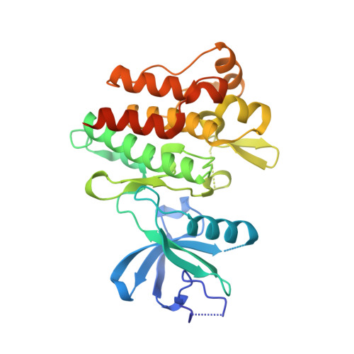
    Allosteric inhibitors of Bcr-Abl have emerged as a novel therapeutic option for the treatment of CML. Using fragment-based screening, a search for novel Abl inhibitors that bind to the myristate pocket was carried out. Here we show that not all myristate ligands are functional inhibitors, but that the conformational state of C-terminal helix_I is a structural determinant for functional activity. We present an NMR-based conformational assay to monitor the conformation of this crucial helix_I and show that myristate ligands that bend helix_I are functional antagonists, whereas ligands that bind to the myristate pocket but do not induce this conformational change are kinase agonists. Activation of c-Abl by allosteric agonists has been confirmed in a biochemical assay.


  • Organizational Affiliation

    Novartis Institutes for Biomedical Research, 4002 Basel, Switzerland. wolfgang.jahnke@novartis.com


Macromolecules
Find similar proteins by:  (by identity cutoff)  |  3D Structure
Entity ID: 1
MoleculeChains Sequence LengthOrganismDetailsImage
Tyrosine-protein kinase ABL1
A, B
293Mus musculusMutation(s): 0 
Gene Names: Abl1Abl
EC: 2.7.10.2
UniProt
Find proteins for P00520 (Mus musculus)
Explore P00520 
Go to UniProtKB:  P00520
Entity Groups  
Sequence Clusters30% Identity50% Identity70% Identity90% Identity95% Identity100% Identity
UniProt GroupP00520
Sequence Annotations
Expand
  • Reference Sequence
Small Molecules
Ligands 3 Unique
IDChains Name / Formula / InChI Key2D Diagram3D Interactions
STI
Query on STI

Download Ideal Coordinates CCD File 
C [auth A],
I [auth B]
4-(4-METHYL-PIPERAZIN-1-YLMETHYL)-N-[4-METHYL-3-(4-PYRIDIN-3-YL-PYRIMIDIN-2-YLAMINO)-PHENYL]-BENZAMIDE
C29 H31 N7 O
KTUFNOKKBVMGRW-UHFFFAOYSA-N
MS9
Query on MS9

Download Ideal Coordinates CCD File 
D [auth A],
J [auth B]
methyl 2-amino-4-chlorobenzoate
C8 H8 Cl N O2
YPSSCICDVDOEAI-UHFFFAOYSA-N
CL
Query on CL

Download Ideal Coordinates CCD File 
E [auth A]
F [auth A]
G [auth A]
H [auth A]
K [auth B]
E [auth A],
F [auth A],
G [auth A],
H [auth A],
K [auth B],
L [auth B]
CHLORIDE ION
Cl
VEXZGXHMUGYJMC-UHFFFAOYSA-M
Binding Affinity Annotations 
IDSourceBinding Affinity
STI BindingDB:  3MS9 Ki: min: 13, max: 7000 (nM) from 5 assay(s)
Kd: min: 1, max: 1.00e+4 (nM) from 35 assay(s)
IC50: min: 1.1, max: 1.00e+4 (nM) from 60 assay(s)
EC50: min: 34, max: 1000 (nM) from 4 assay(s)
MS9 PDBBind:  3MS9 Kd: 6000 (nM) from 1 assay(s)
Experimental Data & Validation

Experimental Data

  • Method: X-RAY DIFFRACTION
  • Resolution: 1.80 Å
  • R-Value Free: 0.234 
  • R-Value Work: 0.186 
  • R-Value Observed: 0.188 
  • Space Group: F 2 2 2
Unit Cell:
Length ( Å )Angle ( ˚ )
a = 115.783α = 90
b = 147.346β = 90
c = 152.833γ = 90
Software Package:
Software NamePurpose
MAR345dtbdata collection
REFMACrefinement
XDSdata reduction
XSCALEdata scaling
REFMACphasing

Structure Validation

View Full Validation Report


Ligand Structure Quality Assessment 


Entry History 

Deposition Data

Revision History  (Full details and data files)

  • Version 1.0: 2010-05-26
    Type: Initial release
  • Version 1.1: 2011-07-13
    Changes: Version format compliance
  • Version 1.2: 2024-02-21
    Changes: Data collection, Database references, Derived calculations, Structure summary