3W56

Structure of a C2 domain


Experimental Data Snapshot

  • Method: X-RAY DIFFRACTION
  • Resolution: 1.60 Å
  • R-Value Free: 0.251 
  • R-Value Work: 0.212 
  • R-Value Observed: 0.214 

wwPDB Validation   3D Report Full Report


This is version 1.2 of the entry. See complete history


Literature

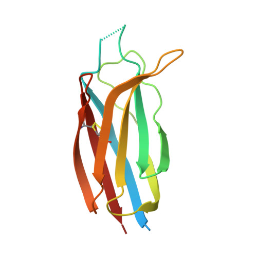
Defining the interaction of perforin with calcium and the phospholipid membrane.

Traore, D.A.Brennan, A.J.Law, R.H.Dogovski, C.Perugini, M.A.Lukoyanova, N.Leung, E.W.Norton, R.S.Lopez, J.A.Browne, K.A.Yagita, H.Lloyd, G.J.Ciccone, A.Verschoor, S.Trapani, J.A.Whisstock, J.C.Voskoboinik, I.

(2013) Biochem J 456: 323-335

  • DOI: https://doi.org/10.1042/BJ20130999
  • Primary Citation of Related Structures:  
    3W56, 3W57

  • PubMed Abstract: 

    Following its secretion from cytotoxic lymphocytes into the immune synapse, perforin binds to target cell membranes through its Ca(2+)-dependent C2 domain. Membrane-bound perforin then forms pores that allow passage of pro-apoptopic granzymes into the target cell. In the present study, structural and biochemical studies reveal that Ca(2+) binding triggers a conformational change in the C2 domain that permits four key hydrophobic residues to interact with the plasma membrane. However, in contrast with previous suggestions, these movements and membrane binding do not trigger irreversible conformational changes in the pore-forming MACPF (membrane attack complex/perforin-like) domain, indicating that subsequent monomer-monomer interactions at the membrane surface are required for perforin pore formation.


  • Organizational Affiliation

    ††Crystallography, Institute of Structural and Molecular Biology, Birkbeck College, Malet Street, London WC1E 7HX, U.K.


Macromolecules
Find similar proteins by:  (by identity cutoff)  |  3D Structure
Entity ID: 1
MoleculeChains Sequence LengthOrganismDetailsImage
C2 domain protein131Scophthalmus maximusMutation(s): 0 
Gene Names: C2P1
UniProt
Find proteins for E2FYL5 (Scophthalmus maximus)
Explore E2FYL5 
Go to UniProtKB:  E2FYL5
Entity Groups  
Sequence Clusters30% Identity50% Identity70% Identity90% Identity95% Identity100% Identity
UniProt GroupE2FYL5
Sequence Annotations
Expand
  • Reference Sequence
Experimental Data & Validation

Experimental Data

  • Method: X-RAY DIFFRACTION
  • Resolution: 1.60 Å
  • R-Value Free: 0.251 
  • R-Value Work: 0.212 
  • R-Value Observed: 0.214 
  • Space Group: I 1 2 1
Unit Cell:
Length ( Å )Angle ( ˚ )
a = 25.348α = 90
b = 55.219β = 91.17
c = 89.746γ = 90
Software Package:
Software NamePurpose
SCALAdata scaling
PHENIXrefinement
PDB_EXTRACTdata extraction

Structure Validation

View Full Validation Report


Entry History 

Deposition Data

Revision History  (Full details and data files)

  • Version 1.0: 2013-10-23
    Type: Initial release
  • Version 1.1: 2014-03-12
    Changes: Database references
  • Version 1.2: 2023-11-08
    Changes: Data collection, Database references, Refinement description