3V6T

Crystal structure of the DNA-bound dHax3, a TAL effector, at 1.85 angstrom


Experimental Data Snapshot

  • Method: X-RAY DIFFRACTION
  • Resolution: 1.85 Å
  • R-Value Free: 0.222 
  • R-Value Work: 0.205 
  • R-Value Observed: 0.206 

wwPDB Validation   3D Report Full Report


This is version 1.1 of the entry. See complete history


Literature

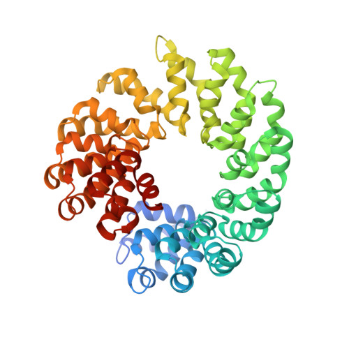
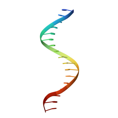
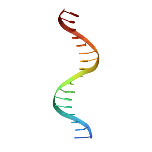
Structural Basis for Sequence-Specific Recognition of DNA by TAL Effectors

Deng, D.Yan, C.Y.Pan, X.J.Mahfouz, M.Wang, J.W.Zhu, J.K.Shi, Y.G.Yan, N.

(2012) Science 

  • DOI: https://doi.org/10.1126/science.1215670
  • Primary Citation of Related Structures:  
    3V6P, 3V6T

  • PubMed Abstract: 

    TAL (transcription activator-like) effectors, secreted by phytopathogenic bacteria, recognize host DNA sequences through a central domain of tandem repeats. Each repeat comprises 33 to 35 conserved amino acids and targets a specific base pair by using two hypervariable residues [known as repeat variable diresidues (RVDs)] at positions 12 and 13. Here, we report the crystal structures of an 11.5-repeat TAL effector in both DNA-free and DNA-bound states. Each TAL repeat comprises two helices connected by a short RVD-containing loop. The 11.5 repeats form a right-handed, superhelical structure that tracks along the sense strand of DNA duplex, with RVDs contacting the major groove. The 12th residue stabilizes the RVD loop, whereas the 13th residue makes a base-specific contact. Understanding DNA recognition by TAL effectors may facilitate rational design of DNA-binding proteins with biotechnological applications.


  • Organizational Affiliation

    State Key Laboratory of Bio-Membrane and Membrane Biotechnology, Tsinghua University, Beijing 100084, China.


Macromolecules

Find similar proteins by:  (by identity cutoff)  |  3D Structure
Entity ID: 1
MoleculeChains Sequence LengthOrganismDetailsImage
dHax3
A, B
499XanthomonasMutation(s): 0 
Entity Groups  
Sequence Clusters30% Identity50% Identity70% Identity90% Identity95% Identity100% Identity
Sequence Annotations
Expand
  • Reference Sequence

Find similar nucleic acids by:  Sequence   |   3D Structure  

Entity ID: 2
MoleculeChains LengthOrganismImage
DNA (5'-D(*TP*GP*TP*CP*CP*CP*TP*TP*TP*AP*TP*CP*TP*CP*TP*CP*T)-3')C [auth G],
E [auth I]
17N/A
Sequence Annotations
Expand
  • Reference Sequence

Find similar nucleic acids by:  Sequence   |   3D Structure  

Entity ID: 3
MoleculeChains LengthOrganismImage
DNA (5'-D(*AP*GP*AP*GP*AP*GP*AP*TP*AP*AP*AP*GP*GP*GP*AP*CP*A)-3')D [auth H],
F [auth J]
17N/A
Sequence Annotations
Expand
  • Reference Sequence
Experimental Data & Validation

Experimental Data

  • Method: X-RAY DIFFRACTION
  • Resolution: 1.85 Å
  • R-Value Free: 0.222 
  • R-Value Work: 0.205 
  • R-Value Observed: 0.206 
  • Space Group: P 1 21 1
Unit Cell:
Length ( Å )Angle ( ˚ )
a = 81.719α = 90
b = 87.679β = 103.04
c = 88.494γ = 90
Software Package:
Software NamePurpose
CBASSdata collection
PHASERphasing
PHENIXrefinement
HKL-2000data reduction
HKL-2000data scaling

Structure Validation

View Full Validation Report


Entry History 

Deposition Data

Revision History  (Full details and data files)

  • Version 1.0: 2012-01-18
    Type: Initial release
  • Version 1.1: 2023-11-08
    Changes: Data collection, Database references, Refinement description