3UGM

Structure of TAL effector PthXo1 bound to its DNA target


Experimental Data Snapshot

  • Method: X-RAY DIFFRACTION
  • Resolution: 3.00 Å
  • R-Value Free: 0.294 
  • R-Value Work: 0.266 

wwPDB Validation   3D Report Full Report


This is version 1.3 of the entry. See complete history


Literature

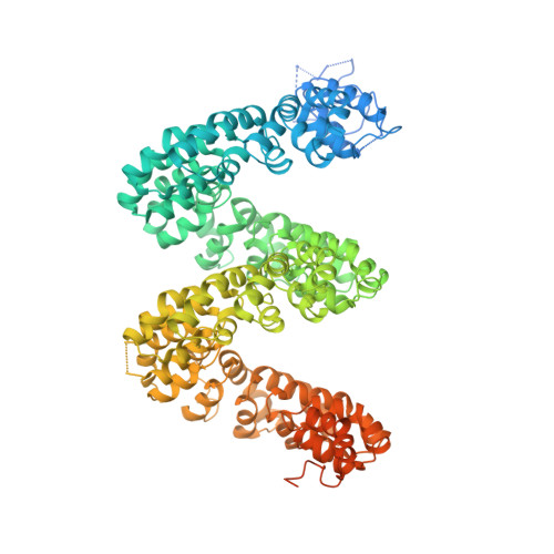
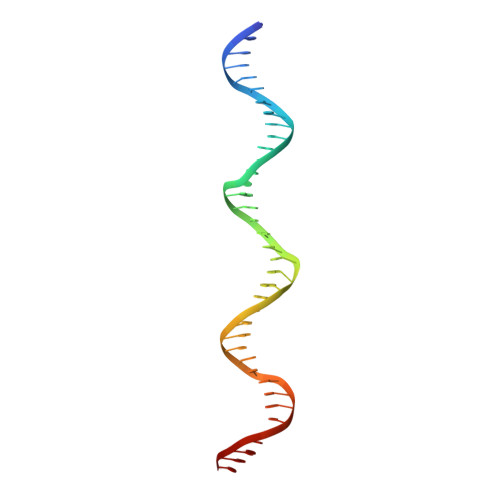
The Crystal Structure of TAL Effector PthXo1 Bound to Its DNA Target.

Mak, A.N.Bradley, P.Cernadas, R.A.Bogdanove, A.J.Stoddard, B.L.

(2012) Science 335: 716-719

  • DOI: https://doi.org/10.1126/science.1216211
  • Primary Citation of Related Structures:  
    3UGM

  • PubMed Abstract: 

    DNA recognition by TAL effectors is mediated by tandem repeats, each 33 to 35 residues in length, that specify nucleotides via unique repeat-variable diresidues (RVDs). The crystal structure of PthXo1 bound to its DNA target was determined by high-throughput computational structure prediction and validated by heavy-atom derivatization. Each repeat forms a left-handed, two-helix bundle that presents an RVD-containing loop to the DNA. The repeats self-associate to form a right-handed superhelix wrapped around the DNA major groove. The first RVD residue forms a stabilizing contact with the protein backbone, while the second makes a base-specific contact to the DNA sense strand. Two degenerate amino-terminal repeats also interact with the DNA. Containing several RVDs and noncanonical associations, the structure illustrates the basis of TAL effector-DNA recognition.


  • Organizational Affiliation

    Division of Basic Sciences, Fred Hutchinson Cancer Research Center, 1100 Fairview Avenue North, A3-025 Seattle, WA 98019, USA.


Macromolecules

Find similar proteins by:  (by identity cutoff)  |  3D Structure
Entity ID: 1
MoleculeChains Sequence LengthOrganismDetailsImage
TAL effector AvrBs3/PthA1,047Xanthomonas oryzae pv. oryzae PXO99AMutation(s): 8 
Gene Names: pthXo1PXO_00227
UniProt
Find proteins for B2SU53 (Xanthomonas oryzae pv. oryzae (strain PXO99A))
Explore B2SU53 
Go to UniProtKB:  B2SU53
Entity Groups  
Sequence Clusters30% Identity50% Identity70% Identity90% Identity95% Identity100% Identity
UniProt GroupB2SU53
Sequence Annotations
Expand
  • Reference Sequence
Find similar nucleic acids by:  (by identity cutoff)  |  3D Structure
Entity ID: 2
MoleculeChains LengthOrganismImage
DNA-138N/A
Sequence Annotations
Expand
  • Reference Sequence
Find similar nucleic acids by:  (by identity cutoff)  |  3D Structure
Entity ID: 3
MoleculeChains LengthOrganismImage
DNA-238N/A
Sequence Annotations
Expand
  • Reference Sequence
Experimental Data & Validation

Experimental Data

  • Method: X-RAY DIFFRACTION
  • Resolution: 3.00 Å
  • R-Value Free: 0.294 
  • R-Value Work: 0.266 
  • Space Group: P 21 21 21
Unit Cell:
Length ( Å )Angle ( ˚ )
a = 95.578α = 90
b = 248.481β = 90
c = 54.646γ = 90
Software Package:
Software NamePurpose
REFMACrefinement
PDB_EXTRACTdata extraction
ADSCdata collection
HKL-2000data reduction
SCALEPACKdata scaling
PHASESphasing
CNSrefinement

Structure Validation

View Full Validation Report


Entry History 

Deposition Data

Revision History  (Full details and data files)

  • Version 1.0: 2012-01-04
    Type: Initial release
  • Version 1.1: 2012-01-25
    Changes: Database references
  • Version 1.2: 2012-02-22
    Changes: Database references
  • Version 1.3: 2024-02-28
    Changes: Data collection, Database references