3OGO

Structure of the GFP:GFP-nanobody complex at 2.8 A resolution in spacegroup P21212


Experimental Data Snapshot

  • Method: X-RAY DIFFRACTION
  • Resolution: 2.80 Å
  • R-Value Free: 0.253 
  • R-Value Work: 0.202 
  • R-Value Observed: 0.204 

wwPDB Validation   3D Report Full Report


This is version 1.3 of the entry. See complete history


Literature

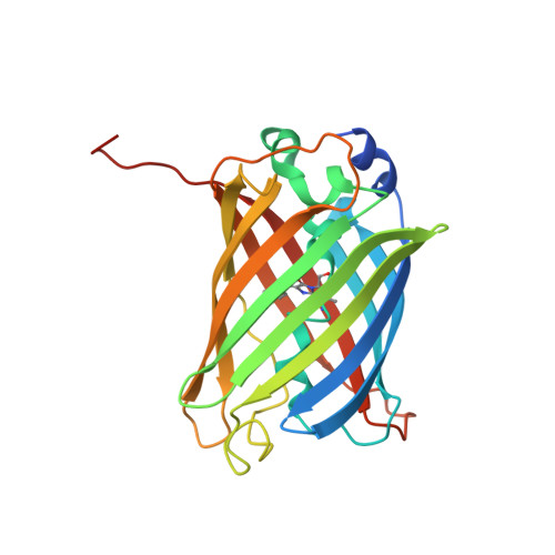
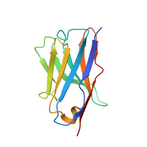
Structural and thermodynamic analysis of the GFP:GFP-nanobody complex.

Kubala, M.H.Kovtun, O.Alexandrov, K.Collins, B.M.

(2010) Protein Sci 19: 2389-2401

  • DOI: https://doi.org/10.1002/pro.519
  • Primary Citation of Related Structures:  
    3OGO

  • PubMed Abstract: 

    The green fluorescent protein (GFP)-nanobody is a single-chain VHH antibody domain developed with specific binding activity against GFP and is emerging as a powerful tool for isolation and cellular engineering of fluorescent protein fusions in many different fields of biological research. Using X-ray crystallography and isothermal titration calorimetry, we determine the molecular details of GFP:GFP-nanobody complex formation and explain the basis of high affinity and at the same time high specificity of protein binding. Although the GFP-nanobody can also bind YFP, it cannot bind the closely related CFP or other fluorescent proteins from the mFruit series. CFP differs from GFP only within the central chromophore and at one surface amino acid position, which lies in the binding interface. Using this information, we have engineered a CFP variant (I146N) that is also able to bind the GFP-nanobody with high affinity, thus extending the toolbox of genetically encoded fluorescent probes that can be isolated using the GFP-nanobody.


  • Organizational Affiliation

    Institute for Molecular Bioscience, University of Queensland, St. Lucia, Queensland 4072, Australia.


Macromolecules
Find similar proteins by:  (by identity cutoff)  |  3D Structure
Entity ID: 1
MoleculeChains Sequence LengthOrganismDetailsImage
Green fluorescent protein
A, B, C, D
247Aequorea victoriaMutation(s): 3 
Gene Names: GFP
UniProt
Find proteins for P42212 (Aequorea victoria)
Explore P42212 
Go to UniProtKB:  P42212
Entity Groups  
Sequence Clusters30% Identity50% Identity70% Identity90% Identity95% Identity100% Identity
UniProt GroupP42212
Sequence Annotations
Expand
  • Reference Sequence
Find similar proteins by:  (by identity cutoff)  |  3D Structure
Entity ID: 2
MoleculeChains Sequence LengthOrganismDetailsImage
GFP-nanobody
E, F, G, H
123Camelus dromedariusMutation(s): 0 
Entity Groups  
Sequence Clusters30% Identity50% Identity70% Identity90% Identity95% Identity100% Identity
Sequence Annotations
Expand
  • Reference Sequence
Small Molecules
Modified Residues  1 Unique
IDChains TypeFormula2D DiagramParent
CRO
Query on CRO
A, B, C, D
L-PEPTIDE LINKINGC15 H17 N3 O5THR, TYR, GLY
Experimental Data & Validation

Experimental Data

  • Method: X-RAY DIFFRACTION
  • Resolution: 2.80 Å
  • R-Value Free: 0.253 
  • R-Value Work: 0.202 
  • R-Value Observed: 0.204 
  • Space Group: P 21 21 2
Unit Cell:
Length ( Å )Angle ( ˚ )
a = 140.1α = 90
b = 147.6β = 90
c = 101.6γ = 90
Software Package:
Software NamePurpose
CrystalCleardata collection
PHASERphasing
PHENIXrefinement
d*TREKdata reduction
d*TREKdata scaling

Structure Validation

View Full Validation Report


Entry History 

Deposition Data

Revision History  (Full details and data files)

  • Version 1.0: 2010-08-25
    Type: Initial release
  • Version 1.1: 2011-07-13
    Changes: Version format compliance
  • Version 1.2: 2021-10-06
    Changes: Advisory, Database references, Derived calculations
  • Version 1.3: 2024-02-21
    Changes: Data collection