3LCK

THE KINASE DOMAIN OF HUMAN LYMPHOCYTE KINASE (LCK), ACTIVATED FORM (AUTO-PHOSPHORYLATED ON TYR394)


Experimental Data Snapshot

  • Method: X-RAY DIFFRACTION
  • Resolution: 1.70 Å
  • R-Value Free: 0.199 
  • R-Value Work: 0.178 
  • R-Value Observed: 0.178 

wwPDB Validation   3D Report Full Report


This is version 1.3 of the entry. See complete history


Literature

Structural basis for activation of human lymphocyte kinase Lck upon tyrosine phosphorylation.

Yamaguchi, H.Hendrickson, W.A.

(1996) Nature 384: 484-489

  • DOI: https://doi.org/10.1038/384484a0
  • Primary Citation of Related Structures:  
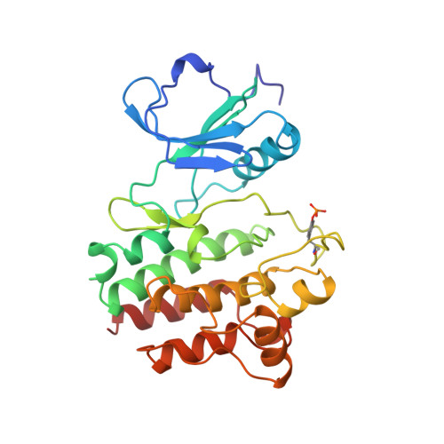
    3LCK

  • PubMed Abstract: 

    Regulation through phosphorylation is a characteristic of signalling pathways and the lymphocyte kinase Lck (p56lck) both performs phosphorylation and is affected by it. Lck is a Src-family tyrosine kinase expressed in T lymphocytes, where it participates in the cellular immune response. Like all Src homologues, it comprises SH3, SH2 and kinase domains. Lck associates through its distinctive amino-terminal segment with the cytoplasmic tails of either T-cell co-receptor, CD4 or CD8-alpha. Activated Lck phosphorylates T-cell receptor zeta-chains, which then recruit the ZAP70 kinase to promote T-cell activation. Lck is activated by autophosphorylation at Tyr 394 in the activation loop and it is inactive when Tyr 505 near the carboxy terminus is phosphorylated and interacts with its own SH2 domain. Here we report the crystal structure of the Lck tyrosine kinase domain (LCKK) in its activated state at 1.7 A resolution. The structure reveals how a phosphoryl group at Tyr 394 generates a competent active site. Comparisons with other kinase structures indicate that tyrosine phophophorylation and ligand binding may in general elicit two distinct hinge-like movements between the kinase subdomains. From modelling studies, we suggest a basis for inhibition by phosphorylation at Tyr 505.


  • Organizational Affiliation

    Department of Biochemistry and Molecular Biophysics, Columbia University, New York 10032, USA.


Macromolecules
Find similar proteins by:  (by identity cutoff)  |  3D Structure
Entity ID: 1
MoleculeChains Sequence LengthOrganismDetailsImage
PROTO-ONCOGENE TYROSINE-PROTEIN KINASE271Homo sapiensMutation(s): 0 
Gene Names: LCK
EC: 2.7.1.112
UniProt & NIH Common Fund Data Resources
Find proteins for P06239 (Homo sapiens)
Explore P06239 
Go to UniProtKB:  P06239
PHAROS:  P06239
GTEx:  ENSG00000182866 
Entity Groups  
Sequence Clusters30% Identity50% Identity70% Identity90% Identity95% Identity100% Identity
UniProt GroupP06239
Sequence Annotations
Expand
  • Reference Sequence
Small Molecules
Ligands 1 Unique
IDChains Name / Formula / InChI Key2D Diagram3D Interactions
SO4
Query on SO4

Download Ideal Coordinates CCD File 
B [auth A]SULFATE ION
O4 S
QAOWNCQODCNURD-UHFFFAOYSA-L
Modified Residues  1 Unique
IDChains TypeFormula2D DiagramParent
PTR
Query on PTR
A
L-PEPTIDE LINKINGC9 H12 N O6 PTYR
Binding Affinity Annotations 
IDSourceBinding Affinity
PTR BindingDB:  3LCK Kd: 4.00e+5 (nM) from 1 assay(s)
Experimental Data & Validation

Experimental Data

  • Method: X-RAY DIFFRACTION
  • Resolution: 1.70 Å
  • R-Value Free: 0.199 
  • R-Value Work: 0.178 
  • R-Value Observed: 0.178 
  • Space Group: P 21 21 21
Unit Cell:
Length ( Å )Angle ( ˚ )
a = 42.04α = 90
b = 73.62β = 90
c = 91.17γ = 90
Software Package:
Software NamePurpose
MADSYSphasing
X-PLORrefinement
DENZOdata reduction
SCALEPACKdata scaling

Structure Validation

View Full Validation Report


Entry History 

Deposition Data

Revision History  (Full details and data files)

  • Version 1.0: 1997-12-03
    Type: Initial release
  • Version 1.1: 2008-03-25
    Changes: Version format compliance
  • Version 1.2: 2011-07-13
    Changes: Version format compliance
  • Version 1.3: 2023-08-09
    Changes: Database references, Derived calculations, Refinement description