3J1S

Structure of adeno-associated virus-2 in complex with neutralizing monoclonal antibody A20


Experimental Data Snapshot

  • Method: ELECTRON MICROSCOPY
  • Resolution: 8.50 Å
  • Aggregation State: PARTICLE 
  • Reconstruction Method: SINGLE PARTICLE 

wwPDB Validation   3D Report Full Report


This is version 1.3 of the entry. See complete history


Literature

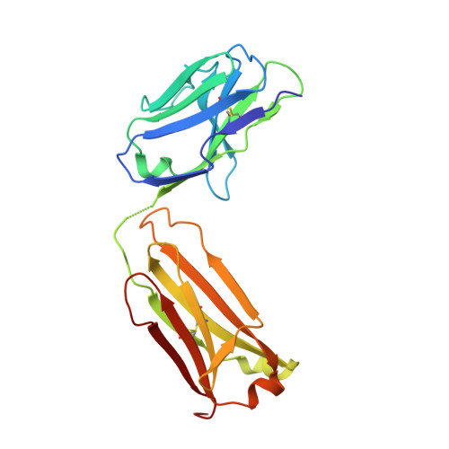
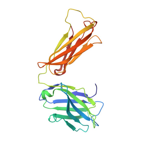
Structure of adeno-associated virus-2 in complex with neutralizing monoclonal antibody A20.

McCraw, D.M.O'Donnell, J.K.Taylor, K.A.Stagg, S.M.Chapman, M.S.

(2012) Virology 431: 40-49

  • DOI: https://doi.org/10.1016/j.virol.2012.05.004
  • Primary Citation of Related Structures:  
    3J1S

  • PubMed Abstract: 

    The use of adeno-associated virus (AAV) as a gene therapy vector is limited by the host neutralizing immune response. The cryo-electron microscopy (EM) structure at 8.5Å resolution is determined for a complex of AAV-2 with the Fab' fragment of monoclonal antibody (MAb) A20, the most extensively characterized AAV MAb. The binding footprint is determined through fitting the cryo-EM reconstruction with a homology model following sequencing of the variable domain, and provides a structural basis for integrating diverse prior epitope mappings. The footprint extends from the previously implicated plateau to the side of the spike, and into the conserved canyon, covering a larger area than anticipated. Comparison with structures of binding and non-binding serotypes indicates that recognition depends on a combination of subtle serotype-specific features. Separation of the neutralizing epitope from the heparan sulfate cell attachment site encourages attempts to develop immune-resistant vectors that can still bind to target cells.


  • Organizational Affiliation

    Department of Biochemistry and Molecular Biology, School of Medicine, Mail code L224, Oregon Health & Science University, 3181 S.W. Sam Jackson Park Road, Portland, OR 97239-3098, USA.


Macromolecules
Find similar proteins by:  (by identity cutoff)  |  3D Structure
Entity ID: 1
MoleculeChains Sequence LengthOrganismDetailsImage
A20 light chainA [auth L]214Mus musculusMutation(s): 0 
Entity Groups  
Sequence Clusters30% Identity50% Identity70% Identity90% Identity95% Identity100% Identity
Sequence Annotations
Expand
  • Reference Sequence
Find similar proteins by:  (by identity cutoff)  |  3D Structure
Entity ID: 2
MoleculeChains Sequence LengthOrganismDetailsImage
A20 heavy chainB [auth H]218Mus musculusMutation(s): 0 
Entity Groups  
Sequence Clusters30% Identity50% Identity70% Identity90% Identity95% Identity100% Identity
Sequence Annotations
Expand
  • Reference Sequence
Find similar proteins by:  (by identity cutoff)  |  3D Structure
Entity ID: 3
MoleculeChains Sequence LengthOrganismDetailsImage
Capsid protein VP1C [auth A]519adeno-associated virus 2Mutation(s): 0 
UniProt
Find proteins for P03135 (Adeno-associated virus 2 (isolate Srivastava/1982))
Explore P03135 
Go to UniProtKB:  P03135
Entity Groups  
Sequence Clusters30% Identity50% Identity70% Identity90% Identity95% Identity100% Identity
UniProt GroupP03135
Sequence Annotations
Expand
  • Reference Sequence
Experimental Data & Validation

Experimental Data

  • Method: ELECTRON MICROSCOPY
  • Resolution: 8.50 Å
  • Aggregation State: PARTICLE 
  • Reconstruction Method: SINGLE PARTICLE 
EM Software:
TaskSoftware PackageVersion
RECONSTRUCTIONAppion
RECONSTRUCTIONEMAN

Structure Validation

View Full Validation Report


Entry History 

Deposition Data

Revision History  (Full details and data files)

  • Version 1.0: 2012-06-06
    Type: Initial release
  • Version 1.1: 2012-06-20
    Changes: Database references
  • Version 1.2: 2012-07-04
    Changes: Database references
  • Version 1.3: 2018-07-18
    Changes: Data collection