2XIG

The structure of the Helicobacter pylori ferric uptake regulator Fur reveals three functional metal binding sites


Experimental Data Snapshot

  • Method: X-RAY DIFFRACTION
  • Resolution: 1.85 Å
  • R-Value Free: 0.234 
  • R-Value Work: 0.198 
  • R-Value Observed: 0.200 

wwPDB Validation   3D Report Full Report


This is version 1.2 of the entry. See complete history


Literature

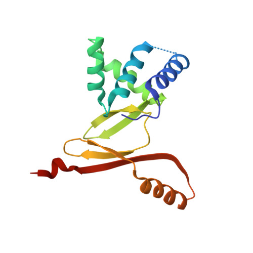
The Structure of the Helicobacter Pylori Ferric Uptake Regulator Fur Reveals Three Functional Metal Binding Sites.

Dian, C.Vitale, S.Leonard, G.A.Bahlawane, C.Fauquant, C.Leduc, D.Muller, C.De Reuse, H.Michaud-Soret, I.Terradot, L.

(2011) Mol Microbiol 79: 1260

  • DOI: https://doi.org/10.1111/j.1365-2958.2010.07517.x
  • Primary Citation of Related Structures:  
    2XIG

  • PubMed Abstract: 

    Fur, the ferric uptake regulator, is a transcription factor that controls iron metabolism in bacteria. Binding of ferrous iron to Fur triggers a conformational change that activates the protein for binding to specific DNA sequences named Fur boxes. In Helicobacter pylori, HpFur is involved in acid response and is important for gastric colonization in model animals. Here we present the crystal structure of a functionally active HpFur mutant (HpFur2M; C78S-C150S) bound to zinc. Although its fold is similar to that of other Fur and Fur-like proteins, the crystal structure of HpFur reveals a unique structured N-terminal extension and an unusual C-terminal helix. The structure also shows three metal binding sites: S1 the structural ZnS₄ site previously characterized biochemically in HpFur and the two zinc sites identified in other Fur proteins. Site-directed mutagenesis and spectroscopy analyses of purified wild-type HpFur and various mutants show that the two metal binding sites common to other Fur proteins can be also metallated by cobalt. DNA protection and circular dichroism experiments demonstrate that, while these two sites influence the affinity of HpFur for DNA, only one is absolutely required for DNA binding and could be responsible for the conformational changes of Fur upon metal binding while the other is a secondary site.


  • Organizational Affiliation

    Structural Biology Group, European Synchrotron Radiation Facility, BP 220 F-38043 Grenoble cedex 9, France.


Macromolecules
Find similar proteins by:  (by identity cutoff)  |  3D Structure
Entity ID: 1
MoleculeChains Sequence LengthOrganismDetailsImage
FERRIC UPTAKE REGULATION PROTEIN
A, B, C, D
150Helicobacter pylori 26695Mutation(s): 2 
UniProt
Find proteins for O25671 (Helicobacter pylori (strain ATCC 700392 / 26695))
Explore O25671 
Go to UniProtKB:  O25671
Entity Groups  
Sequence Clusters30% Identity50% Identity70% Identity90% Identity95% Identity100% Identity
UniProt GroupO25671
Sequence Annotations
Expand
  • Reference Sequence
Small Molecules
Ligands 2 Unique
IDChains Name / Formula / InChI Key2D Diagram3D Interactions
CIT
Query on CIT

Download Ideal Coordinates CCD File 
H [auth B]CITRIC ACID
C6 H8 O7
KRKNYBCHXYNGOX-UHFFFAOYSA-N
ZN
Query on ZN

Download Ideal Coordinates CCD File 
E [auth A]
F [auth A]
G [auth A]
I [auth B]
J [auth B]
E [auth A],
F [auth A],
G [auth A],
I [auth B],
J [auth B],
K [auth B],
L [auth C],
M [auth C],
N [auth C],
O [auth D],
P [auth D],
Q [auth D]
ZINC ION
Zn
PTFCDOFLOPIGGS-UHFFFAOYSA-N
Experimental Data & Validation

Experimental Data

  • Method: X-RAY DIFFRACTION
  • Resolution: 1.85 Å
  • R-Value Free: 0.234 
  • R-Value Work: 0.198 
  • R-Value Observed: 0.200 
  • Space Group: P 1
Unit Cell:
Length ( Å )Angle ( ˚ )
a = 48.15α = 83.32
b = 48.29β = 77.82
c = 72.25γ = 87.65
Software Package:
Software NamePurpose
PHENIXrefinement
MOSFLMdata reduction
SCALAdata scaling
PHASERphasing

Structure Validation

View Full Validation Report


Entry History 

Deposition Data

Revision History  (Full details and data files)

  • Version 1.0: 2011-01-19
    Type: Initial release
  • Version 1.1: 2011-05-08
    Changes: Version format compliance
  • Version 1.2: 2011-07-13
    Changes: Version format compliance