2VAD

Monomeric red fluorescent protein, DsRed.M1


Experimental Data Snapshot

  • Method: X-RAY DIFFRACTION
  • Resolution: 1.59 Å
  • R-Value Free: 0.197 
  • R-Value Work: 0.176 
  • R-Value Observed: 0.177 

wwPDB Validation   3D Report Full Report


This is version 2.1 of the entry. See complete history


Literature

Structural Rearrangements Near the Chromophore Influence the Maturation Speed and Brightness of Dsred Variants.

Strongin, D.E.Bevis, B.Khuong, N.Downing, M.E.Strack, R.L.Sundaram, K.Glick, B.S.Keenan, R.J.

(2007) Protein Eng Des Sel 20: 525

  • DOI: https://doi.org/10.1093/protein/gzm046
  • Primary Citation of Related Structures:  
    2VAD, 2VAE

  • PubMed Abstract: 

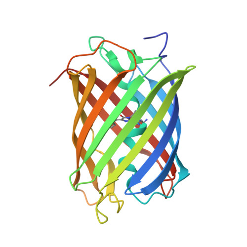
    The red fluorescent protein DsRed has been extensively engineered for use as an in vivo research tool. In fast maturing DsRed variants, the chromophore maturation half-time is approximately 40 min, compared to approximately 12 h for wild-type DsRed. Further, DsRed has been converted from a tetramer into a monomer, a task that entailed mutating approximately 20% of the amino acids. These engineered variants of DsRed have proven extremely valuable for biomedical research, but the structural basis for the improved characteristics has not been thoroughly investigated. Here we present a 1.7 A crystal structure of the fast maturing tetrameric variant DsRed.T4. We also present a biochemical characterization and 1.6 A crystal structure of the monomeric variant DsRed.M1, also known as DsRed-Monomer. Analysis of the crystal structures suggests that rearrangements of Ser69 and Glu215 contribute to fast maturation, and that positioning of the Lys70 side chain modulates fluorescence quantum yield. Despite the 45 mutations in DsRed.M1 relative to wild-type DsRed, there is a root-mean-square deviation of only 0.3 A between the two structures. We propose that novel intramolecular interactions in DsRed.M1 partially compensate for the loss of intermolecular interactions found in the tetramer.


  • Organizational Affiliation

    Department of Molecular Genetics and Cell Biology, University of Chicago, IL 60637, USA.


Macromolecules
Find similar proteins by:  (by identity cutoff)  |  3D Structure
Entity ID: 1
MoleculeChains Sequence LengthOrganismDetailsImage
RED FLUORESCENT PROTEIN223Discosoma sp.Mutation(s): 0 
UniProt
Find proteins for Q9U6Y8 (Discosoma sp.)
Explore Q9U6Y8 
Go to UniProtKB:  Q9U6Y8
Entity Groups  
Sequence Clusters30% Identity50% Identity70% Identity90% Identity95% Identity100% Identity
UniProt GroupQ9U6Y8
Sequence Annotations
Expand
  • Reference Sequence
Small Molecules
Modified Residues  1 Unique
IDChains TypeFormula2D DiagramParent
CRQ
Query on CRQ
A
L-PEPTIDE LINKINGC16 H16 N4 O5GLN, TYR, GLY
Experimental Data & Validation

Experimental Data

  • Method: X-RAY DIFFRACTION
  • Resolution: 1.59 Å
  • R-Value Free: 0.197 
  • R-Value Work: 0.176 
  • R-Value Observed: 0.177 
  • Space Group: P 21 21 21
Unit Cell:
Length ( Å )Angle ( ˚ )
a = 38.892α = 90
b = 62.151β = 90
c = 81.882γ = 90
Software Package:
Software NamePurpose
REFMACrefinement
DENZOdata reduction
SCALEPACKdata scaling
PHASERphasing

Structure Validation

View Full Validation Report


Entry History 

Deposition Data

Revision History  (Full details and data files)

  • Version 1.0: 2007-11-06
    Type: Initial release
  • Version 1.1: 2011-05-08
    Changes: Version format compliance
  • Version 1.2: 2011-07-13
    Changes: Version format compliance
  • Version 1.3: 2019-05-08
    Changes: Data collection, Derived calculations, Experimental preparation, Other
  • Version 2.0: 2023-11-15
    Changes: Atomic model, Data collection, Database references, Derived calculations, Other
  • Version 2.1: 2023-12-13
    Changes: Refinement description