2R17

Functional architecture of the retromer cargo-recognition complex


Experimental Data Snapshot

  • Method: X-RAY DIFFRACTION
  • Resolution: 2.80 Å
  • R-Value Free: 0.268 
  • R-Value Work: 0.216 
  • R-Value Observed: 0.216 

wwPDB Validation   3D Report Full Report


This is version 1.2 of the entry. See complete history


Literature

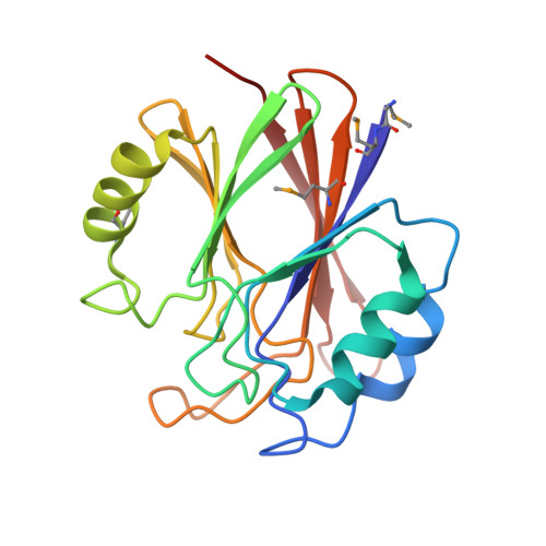
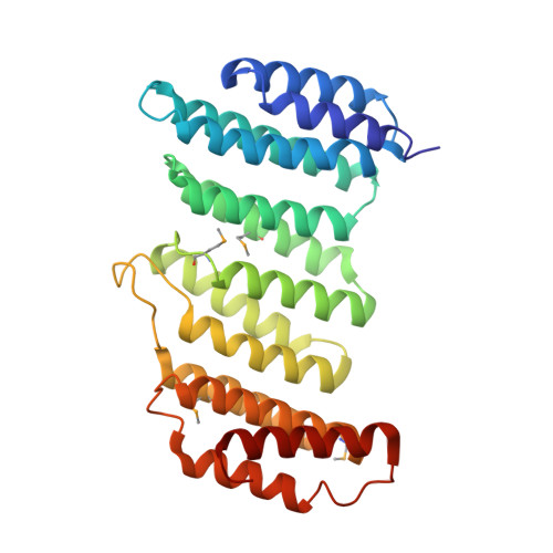
Functional architecture of the retromer cargo-recognition complex.

Hierro, A.Rojas, A.L.Rojas, R.Murthy, N.Effantin, G.Kajava, A.V.Steven, A.C.Bonifacino, J.S.Hurley, J.H.

(2007) Nature 449: 1063-1067

  • DOI: https://doi.org/10.1038/nature06216
  • Primary Citation of Related Structures:  
    2R17

  • PubMed Abstract: 

    The retromer complex is required for the sorting of acid hydrolases to lysosomes, transcytosis of the polymeric immunoglobulin receptor, Wnt gradient formation, iron transporter recycling and processing of the amyloid precursor protein. Human retromer consists of two smaller complexes: the cargo recognition VPS26-VPS29-VPS35 heterotrimer and a membrane-targeting heterodimer or homodimer of SNX1 and/or SNX2 (ref. 13). Here we report the crystal structure of a VPS29-VPS35 subcomplex showing how the metallophosphoesterase-fold subunit VPS29 (refs 14, 15) acts as a scaffold for the carboxy-terminal half of VPS35. VPS35 forms a horseshoe-shaped, right-handed, alpha-helical solenoid, the concave face of which completely covers the metal-binding site of VPS29, whereas the convex face exposes a series of hydrophobic interhelical grooves. Electron microscopy shows that the intact VPS26-VPS29-VPS35 complex is a stick-shaped, flexible structure, approximately 21 nm long. A hybrid structural model derived from crystal structures, electron microscopy, interaction studies and bioinformatics shows that the alpha-solenoid fold extends the full length of VPS35, and that VPS26 is bound at the opposite end from VPS29. This extended structure presents multiple binding sites for the SNX complex and receptor cargo, and appears capable of flexing to conform to curved vesicular membranes.


  • Organizational Affiliation

    Laboratory of Molecular Biology, National Institute of Diabetes and Digestive and Kidney Diseases, National Institutes of Health, US Department of Health and Human Services, Bethesda, Maryland 20892, USA.


Macromolecules
Find similar proteins by:  (by identity cutoff)  |  3D Structure
Entity ID: 1
MoleculeChains Sequence LengthOrganismDetailsImage
Vacuolar protein sorting-associated protein 29
A, B
183Homo sapiensMutation(s): 0 
Gene Names: VPS29
UniProt & NIH Common Fund Data Resources
Find proteins for Q9UBQ0 (Homo sapiens)
Explore Q9UBQ0 
Go to UniProtKB:  Q9UBQ0
PHAROS:  Q9UBQ0
GTEx:  ENSG00000111237 
Entity Groups  
Sequence Clusters30% Identity50% Identity70% Identity90% Identity95% Identity100% Identity
UniProt GroupQ9UBQ0
Sequence Annotations
Expand
  • Reference Sequence
Find similar proteins by:  (by identity cutoff)  |  3D Structure
Entity ID: 2
MoleculeChains Sequence LengthOrganismDetailsImage
Vacuolar protein sorting-associated protein 35
C, D
298Homo sapiensMutation(s): 0 
Gene Names: VPS35MEM3
UniProt & NIH Common Fund Data Resources
Find proteins for Q96QK1 (Homo sapiens)
Explore Q96QK1 
Go to UniProtKB:  Q96QK1
PHAROS:  Q96QK1
GTEx:  ENSG00000069329 
Entity Groups  
Sequence Clusters30% Identity50% Identity70% Identity90% Identity95% Identity100% Identity
UniProt GroupQ96QK1
Sequence Annotations
Expand
  • Reference Sequence
Small Molecules
Modified Residues  1 Unique
IDChains TypeFormula2D DiagramParent
MSE
Query on MSE
A, B
L-PEPTIDE LINKINGC5 H11 N O2 SeMET
Experimental Data & Validation

Experimental Data

  • Method: X-RAY DIFFRACTION
  • Resolution: 2.80 Å
  • R-Value Free: 0.268 
  • R-Value Work: 0.216 
  • R-Value Observed: 0.216 
  • Space Group: P 21 21 21
Unit Cell:
Length ( Å )Angle ( ˚ )
a = 66.25α = 90
b = 128.471β = 90
c = 140.452γ = 90
Software Package:
Software NamePurpose
DENZOdata reduction
SCALEPACKdata scaling
SOLVEphasing
CNSrefinement
PDB_EXTRACTdata extraction
HKL-2000data collection

Structure Validation

View Full Validation Report


Entry History 

Deposition Data

Revision History  (Full details and data files)

  • Version 1.0: 2007-10-30
    Type: Initial release
  • Version 1.1: 2011-07-13
    Changes: Non-polymer description, Version format compliance
  • Version 1.2: 2017-10-25
    Changes: Refinement description