2PX6

Crystal structure of the thioesterase domain of human fatty acid synthase inhibited by Orlistat


Experimental Data Snapshot

  • Method: X-RAY DIFFRACTION
  • Resolution: 2.30 Å
  • R-Value Free: 0.273 
  • R-Value Work: 0.225 
  • R-Value Observed: 0.228 

wwPDB Validation   3D Report Full Report


Ligand Structure Quality Assessment 


This is version 1.3 of the entry. See complete history


Literature

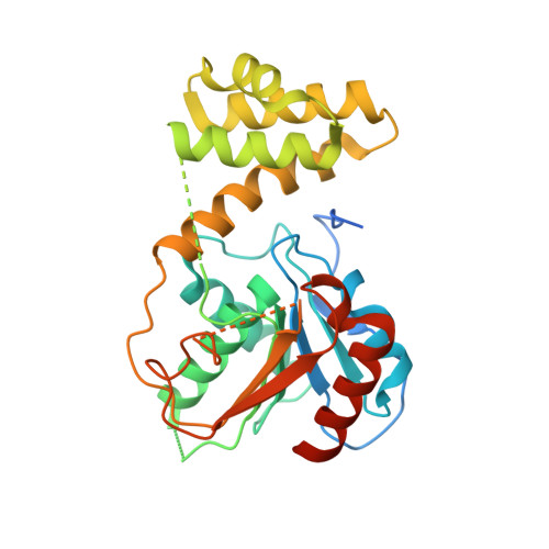
Crystal structure of the thioesterase domain of human fatty acid synthase inhibited by Orlistat.

Pemble, C.W.Johnson, L.C.Kridel, S.J.Lowther, W.T.

(2007) Nat Struct Mol Biol 14: 704-709

  • DOI: https://doi.org/10.1038/nsmb1265
  • Primary Citation of Related Structures:  
    2PX6

  • PubMed Abstract: 

    Human fatty acid synthase (FAS) is uniquely expressed at high levels in many tumor types. Pharmacological inhibition of FAS therefore represents an important therapeutic opportunity. The drug Orlistat, which has been approved by the US Food and Drug Administration, inhibits FAS, induces tumor cell-specific apoptosis and inhibits the growth of prostate tumor xenografts. We determined the 2.3-A-resolution crystal structure of the thioesterase domain of FAS inhibited by Orlistat. Orlistat was captured in the active sites of two thioesterase molecules as a stable acyl-enzyme intermediate and as the hydrolyzed product. The details of these interactions reveal the molecular basis for inhibition and suggest a mechanism for acyl-chain length discrimination during the FAS catalytic cycle. Our findings provide a foundation for the development of new cancer drugs that target FAS.


  • Organizational Affiliation

    Center for Structural Biology and Department of Biochemistry, Wake Forest University School of Medicine, Medical Center Boulevard, Winston-Salem, North Carolina 27157, USA.


Macromolecules
Find similar proteins by:  (by identity cutoff)  |  3D Structure
Entity ID: 1
MoleculeChains Sequence LengthOrganismDetailsImage
Thioesterase domain
A, B
316Homo sapiensMutation(s): 0 
Gene Names: FAS
EC: 2.3.1.85
UniProt & NIH Common Fund Data Resources
Find proteins for P49327 (Homo sapiens)
Explore P49327 
Go to UniProtKB:  P49327
PHAROS:  P49327
GTEx:  ENSG00000169710 
Entity Groups  
Sequence Clusters30% Identity50% Identity70% Identity90% Identity95% Identity100% Identity
UniProt GroupP49327
Sequence Annotations
Expand
  • Reference Sequence
Small Molecules
Ligands 2 Unique
IDChains Name / Formula / InChI Key2D Diagram3D Interactions
DH9
Query on DH9

Download Ideal Coordinates CCD File 
C [auth A],
D [auth B]
(2S,3S,5S)-5-[(N-FORMYL-L-LEUCYL)OXY]-2-HEXYL-3-HYDROXYHEXADECANOIC ACID
C29 H55 N O6
FKUNIADJSAJLGB-FWEHEUNISA-N
DTT
Query on DTT

Download Ideal Coordinates CCD File 
E [auth B]2,3-DIHYDROXY-1,4-DITHIOBUTANE
C4 H10 O2 S2
VHJLVAABSRFDPM-IMJSIDKUSA-N
Experimental Data & Validation

Experimental Data

  • Method: X-RAY DIFFRACTION
  • Resolution: 2.30 Å
  • R-Value Free: 0.273 
  • R-Value Work: 0.225 
  • R-Value Observed: 0.228 
  • Space Group: P 1 21 1
Unit Cell:
Length ( Å )Angle ( ˚ )
a = 41.86α = 90
b = 94.32β = 95.82
c = 69.72γ = 90
Software Package:
Software NamePurpose
REFMACrefinement
CBASSdata collection
d*TREKdata reduction
d*TREKdata scaling
PHASERphasing

Structure Validation

View Full Validation Report


Ligand Structure Quality Assessment 


Entry History 

Deposition Data

Revision History  (Full details and data files)

  • Version 1.0: 2007-07-10
    Type: Initial release
  • Version 1.1: 2008-05-01
    Changes: Version format compliance
  • Version 1.2: 2011-07-13
    Changes: Version format compliance
  • Version 1.3: 2023-08-30
    Changes: Data collection, Database references, Derived calculations, Refinement description