2HZR

Crystal structure of human apolipoprotein D (ApoD)


Experimental Data Snapshot

  • Method: X-RAY DIFFRACTION
  • Resolution: 1.80 Å
  • R-Value Free: 0.250 
  • R-Value Work: 0.202 
  • R-Value Observed: 0.205 

wwPDB Validation   3D Report Full Report


This is version 1.2 of the entry. See complete history


Literature

Structural insight into the dual ligand specificity and mode of high density lipoprotein association of apolipoprotein d.

Eichinger, A.Nasreen, A.Kim, H.J.Skerra, A.

(2007) J Biol Chem 282: 31068-31075

  • DOI: https://doi.org/10.1074/jbc.M703552200
  • Primary Citation of Related Structures:  
    2HZQ, 2HZR

  • PubMed Abstract: 

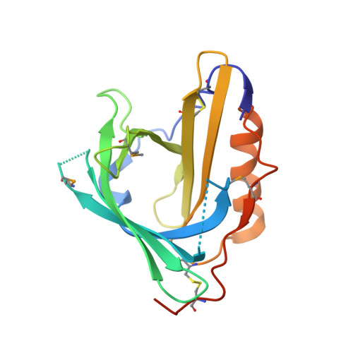
    Human apolipoprotein D (ApoD) occurs in plasma associated with high density lipoprotein. Apart from the involvement in lipid metabolism, its binding activity for progesterone and arachidonic acid plays a role in cancer development and neurological diseases. The crystal structures of free ApoD and its complex with progesterone were determined at 1.8A resolution and reveal a lipocalin fold. The narrow, mainly uncharged pocket within the typical beta-barrel accommodates progesterone with its acetyl side chain oriented toward the bottom. The cavity adopts essentially the same shape in the absence of progesterone and allows complexation of arachidonic acid as another cognate ligand. Three of the four extended loops at the open end of the beta-barrel expose hydrophobic side chains, which is an unusual feature for lipocalins and probably effects association with the high density lipoprotein particle by mediating insertion into the lipid phase. This mechanism is in line with an unpaired Cys residue in the same surface region that can form a disulfide cross-link with apolipoprotein A-II.


  • Organizational Affiliation

    Lehrstuhl für Biologische Chemie, Technische Universität München, 85350 Freising-Weihenstephan, Germany.


Macromolecules
Find similar proteins by:  (by identity cutoff)  |  3D Structure
Entity ID: 1
MoleculeChains Sequence LengthOrganismDetailsImage
Apolipoprotein D174Homo sapiensMutation(s): 10 
Gene Names: APOD
UniProt & NIH Common Fund Data Resources
Find proteins for P05090 (Homo sapiens)
Explore P05090 
Go to UniProtKB:  P05090
PHAROS:  P05090
GTEx:  ENSG00000189058 
Entity Groups  
Sequence Clusters30% Identity50% Identity70% Identity90% Identity95% Identity100% Identity
UniProt GroupP05090
Sequence Annotations
Expand
  • Reference Sequence
Small Molecules
Modified Residues  1 Unique
IDChains TypeFormula2D DiagramParent
MSE
Query on MSE
A
L-PEPTIDE LINKINGC5 H11 N O2 SeMET
Experimental Data & Validation

Experimental Data

  • Method: X-RAY DIFFRACTION
  • Resolution: 1.80 Å
  • R-Value Free: 0.250 
  • R-Value Work: 0.202 
  • R-Value Observed: 0.205 
  • Space Group: P 43 21 2
Unit Cell:
Length ( Å )Angle ( ˚ )
a = 49.247α = 90
b = 49.247β = 90
c = 143.486γ = 90
Software Package:
Software NamePurpose
SCALAdata scaling
SHARPphasing
REFMACrefinement
PDB_EXTRACTdata extraction
ADSCdata collection
MOSFLMdata reduction

Structure Validation

View Full Validation Report


Entry History 

Deposition Data

Revision History  (Full details and data files)

  • Version 1.0: 2007-08-14
    Type: Initial release
  • Version 1.1: 2011-07-13
    Changes: Version format compliance
  • Version 1.2: 2021-10-20
    Changes: Database references, Derived calculations