2ERK

PHOSPHORYLATED MAP KINASE ERK2


Experimental Data Snapshot

  • Method: X-RAY DIFFRACTION
  • Resolution: 2.40 Å
  • R-Value Free: 0.264 
  • R-Value Work: 0.192 
  • R-Value Observed: 0.192 

wwPDB Validation   3D Report Full Report


This is version 1.3 of the entry. See complete history


Literature

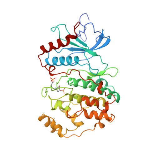
Activation mechanism of the MAP kinase ERK2 by dual phosphorylation.

Canagarajah, B.J.Khokhlatchev, A.Cobb, M.H.Goldsmith, E.J.

(1997) Cell 90: 859-869

  • DOI: https://doi.org/10.1016/s0092-8674(00)80351-7
  • Primary Citation of Related Structures:  
    2ERK

  • PubMed Abstract: 

    The structure of the active form of the MAP kinase ERK2 has been solved, phosphorylated on a threonine and a tyrosine residue within the phosphorylation lip. The lip is refolded, bringing the phosphothreonine and phosphotyrosine into alignment with surface arginine-rich binding sites. Conformational changes occur in the lip and neighboring structures, including the P+1 site, the MAP kinase insertion, the C-terminal extension, and helix C. Domain rotation and remodeling of the proline-directed P+1 specificity pocket account for the activation. The conformation of the P+1 pocket is similar to a second proline-directed kinase, CDK2-CyclinA, thus permitting the origin of this specificity to be defined. Conformational changes outside the lip provide loci at which the state of phosphorylation can be felt by other cellular components.


  • Organizational Affiliation

    Department of Biochemistry, The University of Texas Southwestern Medical Center at Dallas, 75235-9050, USA.


Macromolecules
Find similar proteins by:  (by identity cutoff)  |  3D Structure
Entity ID: 1
MoleculeChains Sequence LengthOrganismDetailsImage
EXTRACELLULAR SIGNAL-REGULATED KINASE 2365Rattus norvegicusMutation(s): 2 
Gene Names: ERK2
EC: 2.7.1
UniProt
Find proteins for P63086 (Rattus norvegicus)
Explore P63086 
Go to UniProtKB:  P63086
Entity Groups  
Sequence Clusters30% Identity50% Identity70% Identity90% Identity95% Identity100% Identity
UniProt GroupP63086
Sequence Annotations
Expand
  • Reference Sequence
Small Molecules
Modified Residues  2 Unique
IDChains TypeFormula2D DiagramParent
PTR
Query on PTR
A
L-PEPTIDE LINKINGC9 H12 N O6 PTYR
TPO
Query on TPO
A
L-PEPTIDE LINKINGC4 H10 N O6 PTHR
Experimental Data & Validation

Experimental Data

  • Method: X-RAY DIFFRACTION
  • Resolution: 2.40 Å
  • R-Value Free: 0.264 
  • R-Value Work: 0.192 
  • R-Value Observed: 0.192 
  • Space Group: P 41 21 2
Unit Cell:
Length ( Å )Angle ( ˚ )
a = 92.5α = 90
b = 92.5β = 90
c = 103γ = 90
Software Package:
Software NamePurpose
X-PLORmodel building
X-PLORrefinement
DENZOdata reduction
SCALEPACKdata scaling
X-PLORphasing

Structure Validation

View Full Validation Report


Entry History 

Deposition Data

Revision History  (Full details and data files)

  • Version 1.0: 1998-07-01
    Type: Initial release
  • Version 1.1: 2008-03-24
    Changes: Version format compliance
  • Version 1.2: 2011-07-13
    Changes: Version format compliance
  • Version 1.3: 2023-08-09
    Changes: Database references, Derived calculations, Other, Refinement description