


The 3.3 A Crystallographic Structure of Restriction Endonuclease Eco RI in the Absence of DNA
Chandrasekhar, K., Horvath, M.M., Samudzi, C., Choi, J., Rosenberg, J.M.To be published.
Experimental Data Snapshot
wwPDB Validation 3D Report Full Report

Entity ID: 1 | |||||
|---|---|---|---|---|---|
| Molecule | Chains | Sequence Length | Organism | Details | Image |
| PROTEIN (ECO RI ENDONUCLEASE) | 276 | Escherichia coli | Mutation(s): 0 EC: 3.1.21.4 | 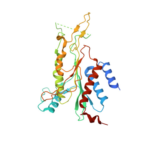 | |
UniProt | |||||
Find proteins for P00642 (Escherichia coli) Explore P00642 Go to UniProtKB: P00642 | |||||
Entity Groups | |||||
| Sequence Clusters | 30% Identity50% Identity70% Identity90% Identity95% Identity100% Identity | ||||
| UniProt Group | P00642 | ||||
Sequence AnnotationsExpand | |||||
| |||||
| Length ( Å ) | Angle ( ˚ ) |
|---|---|
| a = 208.68 | α = 90 |
| b = 127.35 | β = 98.57 |
| c = 49.87 | γ = 90 |
| Software Name | Purpose |
|---|---|
| MERLOT | phasing |
| X-PLOR | refinement |
| X-GEN | data reduction |
| X-GEN | data scaling |
RCSB PDB (citation) is hosted by
RCSB PDB is a member of the

