Solution NMR Structure Investigation for Releasing Mechanism of Neocarzinostatin Chromophore from the Holoprotein
Takashima, H., Yoshida, T., Ishino, T., Hasuda, K., Ohkubo, T., Kobayashi, Y.(2005) J Biol Chem 280: 11340-11346
- PubMed: 15640161
- DOI: https://doi.org/10.1074/jbc.M411579200
- Primary Citation of Related Structures:
1O5P - PubMed Abstract:
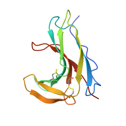
Holo-neocarzinostatin (holo-NCS) is a complex protein carrying the anti-tumor active enediyne ring chromophore by a scaffold consisting of an immunoglobulin-like seven-stranded anti-parallel beta-barrel. Because of the labile chromophore reflecting its extremely strong DNA cleavage activity and complete stabilization in the complex, holo-NCS has attracted much attention in clinical use as well as for drug delivery systems. Despite many structural analyses for holo-NCS, the chromophore-releasing mechanism to trigger prompt attacks on the target DNA is still unclear. We determined the three-dimensional structure of the protein and the internal motion by multinuclear NMR to investigate the releasing mechanism. The internal motion studied by 13C NMR methine relaxation experiments showed that the complex has a rigid structure for its loops as well as the beta-barrel in aqueous solution. This agrees with the refined NMR solution structure, which has good convergence in the loop regions. We also showed that the chromophore displayed a similar internal motion as the protein moiety. The structural comparison between the refined solution structure and x-ray crystal structure indicated characteristic differences. Based on the findings, we proposed the chromophore-releasing mechanism by a three-state equilibrium, which sufficiently describes both the strong binding and the prompt releasing of the chromophore. We demonstrated that we could bridge the dynamic properties and the static structure features with simple kinetic assumptions to solve the biochemical function.
Organizational Affiliation:
Informatics and Knowledge Management at Novartis Institutes for BioMedical Research, Novartis, Ohkubo 8, Tsukuba, Ibaraki 300-2611, Japan.