1M2X

Crystal Structure of the metallo-beta-lactamase BlaB of Chryseobacterium meningosepticum in complex with the inhibitor D-captopril


Experimental Data Snapshot

  • Method: X-RAY DIFFRACTION
  • Resolution: 1.50 Å
  • R-Value Free: 0.208 
  • R-Value Work: 0.190 

wwPDB Validation   3D Report Full Report


Ligand Structure Quality Assessment 


This is version 1.3 of the entry. See complete history


Literature

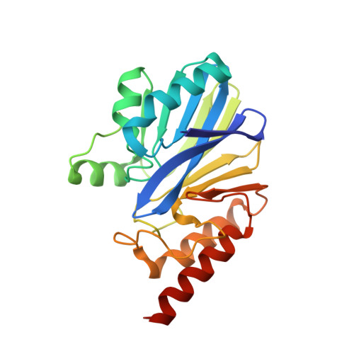
The 1.5 A structure of Chryseobacterium meningosepticum Zn-beta-lactamase in complex with the inhibitor, D-captopril

Garcia-Saez, I.Hopkins, J.Papamicael, C.Franceschini, N.Amicosante, G.Rossolini, G.M.Galleni, M.Frere, J.M.Dideberg, O.

(2003) J Biol Chem 278: 23868-23873

  • DOI: https://doi.org/10.1074/jbc.M301062200
  • Primary Citation of Related Structures:  
    1M2X

  • PubMed Abstract: 

    The crystal structure of the class-B beta-lactamase, BlaB, from the pathogenic bacterium, Chryseobacterium meningosepticum, in complex with the inhibitor, d-captopril, has been solved at 1.5-A resolution. The enzyme has the typical alphabeta/betaalpha metallo-beta-lactamase fold and the characteristic two metal binding sites of members of the subclass B1, in which two Zn2+ ions were identified. d-Captopril, a diastereoisomer of the commercial drug, captopril, acts as an inhibitor by displacing the catalytic hydroxyl ion required for antibiotic hydrolysis and intercalating its sulfhydryl group between the two Zn2+ ions. Interestingly, d-captopril is located on one side of the active site cleft. The x-ray structure of the complex of the closely related enzyme, IMP-1, with a mercaptocarboxylate inhibitor, which also contains a sulfhydryl group bound to the two Zn2+ ions, shows the ligand to be located on the opposite side of the active site cleft. A molecule generated by fusion of these two inhibitors would cover the entire cleft, suggesting an interesting approach to the design of highly specific inhibitors.


  • Organizational Affiliation

    Laboratoire de Cristallographie Macromoléculaire, Institut de Biologie Structurale Jean-Pierre Ebel (CNRS-Commissariat à l'Energie Atomique, Saclay, France.


Macromolecules
Find similar proteins by:  (by identity cutoff)  |  3D Structure
Entity ID: 1
MoleculeChains Sequence LengthOrganismDetailsImage
class B carbapenemase BlaB-1
A, B, C, D
223Elizabethkingia meningosepticaMutation(s): 0 
Gene Names: blaB
EC: 3.5.2.6
UniProt
Find proteins for O08498 (Elizabethkingia meningoseptica)
Explore O08498 
Go to UniProtKB:  O08498
Entity Groups  
Sequence Clusters30% Identity50% Identity70% Identity90% Identity95% Identity100% Identity
UniProt GroupO08498
Sequence Annotations
Expand
  • Reference Sequence
Small Molecules
Ligands 4 Unique
IDChains Name / Formula / InChI Key2D Diagram3D Interactions
MCO
Query on MCO

Download Ideal Coordinates CCD File 
I [auth A],
N [auth B],
S [auth C],
X [auth D]
1-(3-MERCAPTO-2-METHYL-PROPIONYL)-PYRROLIDINE-2-CARBOXYLIC ACID
C9 H15 N O3 S
FAKRSMQSSFJEIM-RNFRBKRXSA-N
GOL
Query on GOL

Download Ideal Coordinates CCD File 
J [auth A],
O [auth B],
T [auth C],
Y [auth D]
GLYCEROL
C3 H8 O3
PEDCQBHIVMGVHV-UHFFFAOYSA-N
ZN
Query on ZN

Download Ideal Coordinates CCD File 
F [auth A]
G [auth A]
H [auth A]
K [auth B]
L [auth B]
F [auth A],
G [auth A],
H [auth A],
K [auth B],
L [auth B],
M [auth B],
P [auth C],
Q [auth C],
R [auth C],
U [auth D],
V [auth D],
W [auth D]
ZINC ION
Zn
PTFCDOFLOPIGGS-UHFFFAOYSA-N
NA
Query on NA

Download Ideal Coordinates CCD File 
E [auth A]SODIUM ION
Na
FKNQFGJONOIPTF-UHFFFAOYSA-N
Binding Affinity Annotations 
IDSourceBinding Affinity
MCO Binding MOAD:  1M2X Ki: 8.50e+4 (nM) from 1 assay(s)
PDBBind:  1M2X Ki: 7.00e+4 (nM) from 1 assay(s)
Experimental Data & Validation

Experimental Data

  • Method: X-RAY DIFFRACTION
  • Resolution: 1.50 Å
  • R-Value Free: 0.208 
  • R-Value Work: 0.190 
  • Space Group: H 3
Unit Cell:
Length ( Å )Angle ( ˚ )
a = 159.685α = 90
b = 159.685β = 90
c = 97.815γ = 120
Software Package:
Software NamePurpose
DENZOdata reduction
SCALAdata scaling
AMoREphasing
CNSrefinement
CCP4data scaling

Structure Validation

View Full Validation Report


Ligand Structure Quality Assessment 


Entry History 

Deposition Data

Revision History  (Full details and data files)

  • Version 1.0: 2003-07-29
    Type: Initial release
  • Version 1.1: 2008-04-28
    Changes: Version format compliance
  • Version 1.2: 2011-07-13
    Changes: Non-polymer description, Version format compliance
  • Version 1.3: 2024-02-14
    Changes: Data collection, Database references, Derived calculations, Refinement description