1J42

Crystal Structure of Human DJ-1


Experimental Data Snapshot

  • Method: X-RAY DIFFRACTION
  • Resolution: 2.50 Å
  • R-Value Free: 0.249 
  • R-Value Work: 0.228 
  • R-Value Observed: 0.228 

wwPDB Validation   3D Report Full Report


This is version 1.4 of the entry. See complete history


Literature

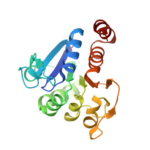
Crystal structures of human DJ-1 and Escherichia coli Hsp31, which share an evolutionarily conserved domain.

Lee, S.J.Kim, S.J.Kim, I.K.Ko, J.Jeong, C.S.Kim, G.H.Park, C.Kang, S.O.Suh, P.G.Lee, H.S.Cha, S.S.

(2003) J Biol Chem 278: 44552-44559

  • DOI: https://doi.org/10.1074/jbc.M304517200
  • Primary Citation of Related Structures:  
    1IZY, 1IZZ, 1J42

  • PubMed Abstract: 

    Human DJ-1 and Escherichia coli Hsp31 belong to ThiJ/PfpI family, whose members contain a conserved domain. DJ-1 is associated with autosomal recessive early onset parkinsonism and Hsp31 is a molecular chaperone. Structural comparisons between DJ-1, Hsp31, and an Archaea protease, a member of ThiJ/PfpI family, lead to the identification of the chaperone activity of DJ-1 and the proteolytic activity of Hsp31. Moreover, the comparisons provide insights into how the functional diversity is realized in proteins that share an evolutionarily conserved domain. On the basis of the chaperone activity the possible role of DJ-1 in the pathogenesis of Parkinson's disease is discussed.


  • Organizational Affiliation

    Beamline Division, Pohang Accelerator Laboratory, Pohang, 790-784, Kyungbuk, Republic of Korea.


Macromolecules
Find similar proteins by:  (by identity cutoff)  |  3D Structure
Entity ID: 1
MoleculeChains Sequence LengthOrganismDetailsImage
RNA-binding protein regulatory subunit189Homo sapiensMutation(s): 0 
UniProt & NIH Common Fund Data Resources
Find proteins for Q99497 (Homo sapiens)
Explore Q99497 
Go to UniProtKB:  Q99497
PHAROS:  Q99497
GTEx:  ENSG00000116288 
Entity Groups  
Sequence Clusters30% Identity50% Identity70% Identity90% Identity95% Identity100% Identity
UniProt GroupQ99497
Sequence Annotations
Expand
  • Reference Sequence
Experimental Data & Validation

Experimental Data

  • Method: X-RAY DIFFRACTION
  • Resolution: 2.50 Å
  • R-Value Free: 0.249 
  • R-Value Work: 0.228 
  • R-Value Observed: 0.228 
  • Space Group: P 31 2 1
Unit Cell:
Length ( Å )Angle ( ˚ )
a = 75.075α = 90
b = 75.075β = 90
c = 75.158γ = 120
Software Package:
Software NamePurpose
CNSrefinement
SCALEPACKdata scaling
CNSphasing

Structure Validation

View Full Validation Report


Entry History 

Deposition Data

  • Released Date: 2004-02-03 
  • Deposition Author(s): Cha, S.S.

Revision History  (Full details and data files)

  • Version 1.0: 2004-02-03
    Type: Initial release
  • Version 1.1: 2008-04-27
    Changes: Version format compliance
  • Version 1.2: 2011-07-13
    Changes: Derived calculations, Version format compliance
  • Version 1.3: 2017-10-04
    Changes: Refinement description
  • Version 1.4: 2023-12-27
    Changes: Data collection, Database references