1GNH

HUMAN C-REACTIVE PROTEIN


Experimental Data Snapshot

  • Method: X-RAY DIFFRACTION
  • Resolution: 3.00 Å
  • R-Value Free: 0.273 
  • R-Value Work: 0.239 
  • R-Value Observed: 0.239 

wwPDB Validation   3D Report Full Report


This is version 1.2 of the entry. See complete history


Literature

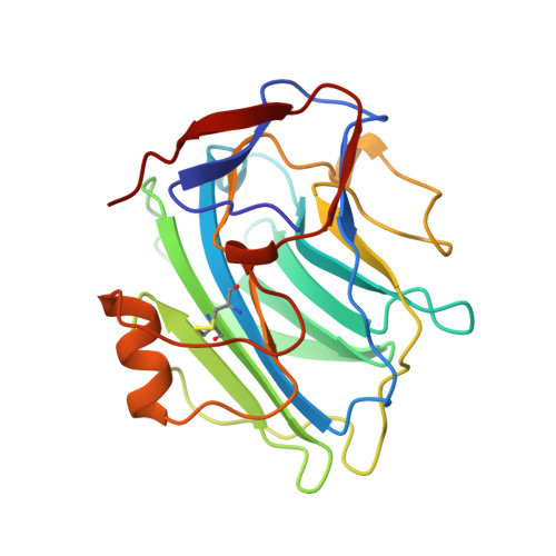
Three dimensional structure of human C-reactive protein.

Shrive, A.K.Cheetham, G.M.Holden, D.Myles, D.A.Turnell, W.G.Volanakis, J.E.Pepys, M.B.Bloomer, A.C.Greenhough, T.J.

(1996) Nat Struct Biol 3: 346-354

  • DOI: https://doi.org/10.1038/nsb0496-346
  • Primary Citation of Related Structures:  
    1GNH

  • PubMed Abstract: 

    The structure of the classical acute phase reactant human C-reactive protein provides evidence that phosphocholine binding is mediated through calcium and a hydrophobic pocket centred on Phe 66. The residue Glu 81 is suitably positioned to interact with the choline group. A cleft on the pentameric face opposite to that containing the calcium site may have an important functional role. The structure provides insights into the molecular mechanisms by which this highly conserved plasma protein, for which no polymorphism or deficiency state is known, may exert its biological role.


  • Organizational Affiliation

    Department of Physics, Keele University, Keele, UK.


Macromolecules
Find similar proteins by:  (by identity cutoff)  |  3D Structure
Entity ID: 1
MoleculeChains Sequence LengthOrganismDetailsImage
C-REACTIVE PROTEIN
A, B, C, D, E
A, B, C, D, E, F, G, H, I, J
206Homo sapiensMutation(s): 0 
UniProt & NIH Common Fund Data Resources
Find proteins for P02741 (Homo sapiens)
Explore P02741 
Go to UniProtKB:  P02741
PHAROS:  P02741
GTEx:  ENSG00000132693 
Entity Groups  
Sequence Clusters30% Identity50% Identity70% Identity90% Identity95% Identity100% Identity
UniProt GroupP02741
Sequence Annotations
Expand
  • Reference Sequence
Small Molecules
Ligands 1 Unique
IDChains Name / Formula / InChI Key2D Diagram3D Interactions
CA
Query on CA

Download Ideal Coordinates CCD File 
K [auth A]
L [auth A]
M [auth B]
N [auth B]
O [auth C]
K [auth A],
L [auth A],
M [auth B],
N [auth B],
O [auth C],
P [auth C],
Q [auth E],
R [auth E],
S [auth F],
T [auth F],
U [auth G],
V [auth G],
W [auth H],
X [auth H],
Y [auth I],
Z [auth I]
CALCIUM ION
Ca
BHPQYMZQTOCNFJ-UHFFFAOYSA-N
Experimental Data & Validation

Experimental Data

  • Method: X-RAY DIFFRACTION
  • Resolution: 3.00 Å
  • R-Value Free: 0.273 
  • R-Value Work: 0.239 
  • R-Value Observed: 0.239 
  • Space Group: P 21 21 21
Unit Cell:
Length ( Å )Angle ( ˚ )
a = 101.06α = 90
b = 154.25β = 90
c = 242.93γ = 90
Software Package:
Software NamePurpose
DENZOdata reduction
CCP4data collection
X-PLORmodel building
X-PLORrefinement
CCP4data reduction
X-PLORphasing

Structure Validation

View Full Validation Report


Entry History 

Deposition Data

Revision History  (Full details and data files)

  • Version 1.0: 1997-01-27
    Type: Initial release
  • Version 1.1: 2008-03-24
    Changes: Version format compliance
  • Version 1.2: 2011-07-13
    Changes: Version format compliance