1BM0

CRYSTAL STRUCTURE OF HUMAN SERUM ALBUMIN


Experimental Data Snapshot

  • Method: X-RAY DIFFRACTION
  • Resolution: 2.50 Å
  • R-Value Free: 0.292 
  • R-Value Work: 0.207 
  • R-Value Observed: 0.207 

wwPDB Validation   3D Report Full Report


This is version 1.3 of the entry. See complete history


Literature

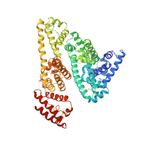
Crystal structure of human serum albumin at 2.5 A resolution.

Sugio, S.Kashima, A.Mochizuki, S.Noda, M.Kobayashi, K.

(1999) Protein Eng 12: 439-446

  • DOI: https://doi.org/10.1093/protein/12.6.439
  • Primary Citation of Related Structures:  
    1AO6, 1BM0

  • PubMed Abstract: 

    A new triclinic crystal form of human serum albumin (HSA), derived either from pool plasma (pHSA) or from a Pichia pastoris expression system (rHSA), was obtained from polyethylene glycol 4000 solution. Three-dimensional structures of pHSA and rHSA were determined at 2.5 A resolution from the new triclinic crystal form by molecular replacement, using atomic coordinates derived from a multiple isomorphous replacement work with a known tetragonal crystal form. The structures of pHSA and rHSA are virtually identical, with an r.m. s. deviation of 0.24 A for all Calpha atoms. The two HSA molecules involved in the asymmetric unit are related by a strict local twofold symmetry such that the Calpha atoms of the two molecules can be superimposed with an r.m.s. deviation of 0.28 A in pHSA. Cys34 is the only cysteine with a free sulfhydryl group which does not participate in a disulfide linkage with any external ligand. Domains II and III both have a pocket formed mostly of hydrophobic and positively charged residues and in which a very wide range of compounds may be accommodated. Three tentative binding sites for long-chain fatty acids, each with different surroundings, are located at the surface of each domain.


  • Organizational Affiliation

    Osaka Laboratories, Yoshitomi Pharmaceutical Industries Ltd,2-25-1, Shodai-Ohtani, Hirakata, Osaka 573-1153, Japan.


Macromolecules
Find similar proteins by:  (by identity cutoff)  |  3D Structure
Entity ID: 1
MoleculeChains Sequence LengthOrganismDetailsImage
SERUM ALBUMIN
A, B
585Homo sapiensMutation(s): 0 
UniProt & NIH Common Fund Data Resources
Find proteins for P02768 (Homo sapiens)
Explore P02768 
Go to UniProtKB:  P02768
PHAROS:  P02768
GTEx:  ENSG00000163631 
Entity Groups  
Sequence Clusters30% Identity50% Identity70% Identity90% Identity95% Identity100% Identity
UniProt GroupP02768
Sequence Annotations
Expand
  • Reference Sequence
Experimental Data & Validation

Experimental Data

  • Method: X-RAY DIFFRACTION
  • Resolution: 2.50 Å
  • R-Value Free: 0.292 
  • R-Value Work: 0.207 
  • R-Value Observed: 0.207 
  • Space Group: P 1
Unit Cell:
Length ( Å )Angle ( ˚ )
a = 59.36α = 91.04
b = 96.89β = 103.4
c = 59.24γ = 74.94
Software Package:
Software NamePurpose
CONTROLdata collection
PROCESSdata reduction
X-PLORmodel building
X-PLORrefinement
CONTROLdata reduction
PROCESSdata scaling
X-PLORphasing

Structure Validation

View Full Validation Report


Entry History 

Deposition Data

Revision History  (Full details and data files)

  • Version 1.0: 1999-07-28
    Type: Initial release
  • Version 1.1: 2008-03-24
    Changes: Version format compliance
  • Version 1.2: 2011-07-13
    Changes: Version format compliance
  • Version 1.3: 2023-08-02
    Changes: Database references, Other, Refinement description